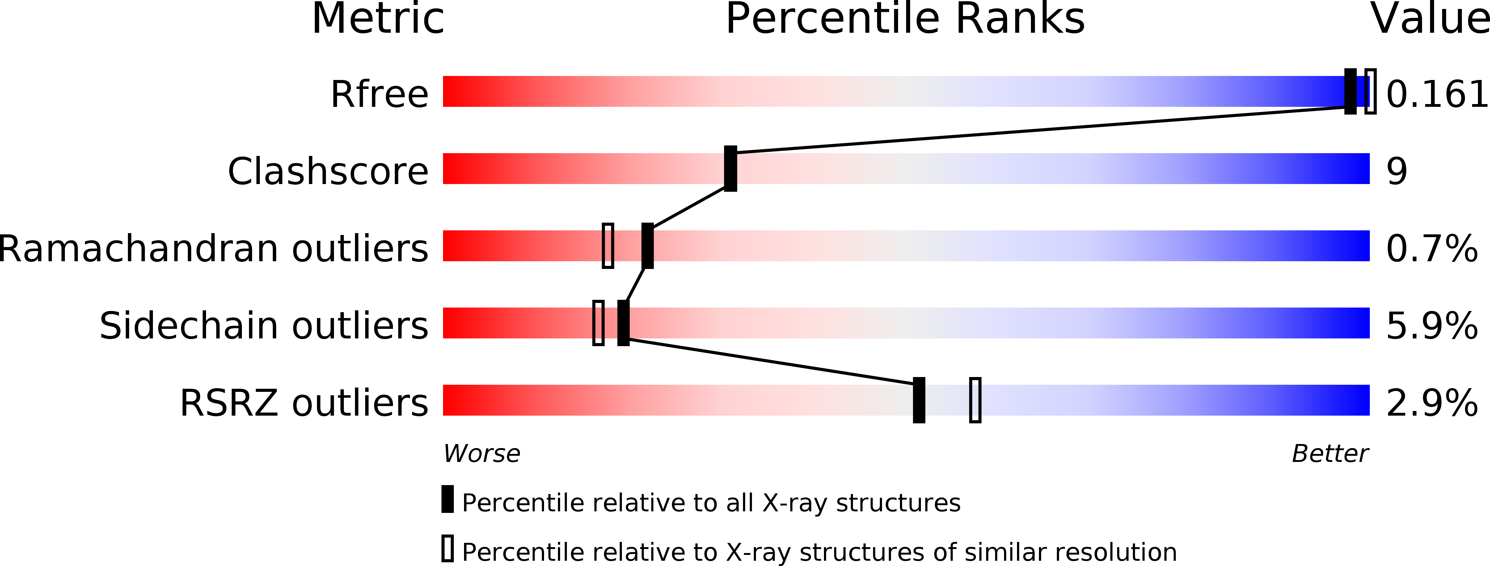
2DU2 image
Deposition Date 2006-07-19
Release Date 2006-12-05
Last Version Date 2023-10-25
Entry Detail
PDB ID:
2DU2
Keywords:
Title:
Crystal Structure Analysis of the L-Lactate Oxidase
Biological Source:
Source Organism:
Method Details:
Experimental Method:
Resolution:
2.10 Å
R-Value Free:
0.19
R-Value Work:
0.15
R-Value Observed:
0.16
Space Group:
I 4 2 2
Macromolecular Entities
Structures with similar UniProt ID
Protein Blast
Polymer Type:polypeptide(L)
Molecule:Lactate oxidase
Chain IDs:A, B, C, D
Chain Length:374
Number of Molecules:4
Biological Source:Aerococcus viridans
Ligand Molecules
Primary Citation
The crystal structure of L-lactate oxidase from Aerococcus viridans at 2.1A resolution reveals the mechanism of strict substrate recognition
Biochem.Biophys.Res.Commun. 350 249 256 (2006)
PMID: 17007814 DOI: 10.1016/j.bbrc.2006.09.025

Abstact

L-Lactate oxidase (LOX) from Aerococcus viridans is a member of the alpha-hydroxyacid-oxidase flavoenzyme family. We have determined the three-dimensional structure of LOX and revealed the mechanism of substrate recognition. The LOX monomer structure has a typical alpha(8)/beta(8) motif commonly found in other flavin family proteins. A related enzyme, glycolate oxidase, catalyzes the oxidation of glycolate rather than lactate. Comparison of the two enzyme structures highlights the importance of five residues around the FMN prosthetic group of LOX, which act synergistically to discriminate between the l/d configurations of lactate. X-ray crystallography of LOX gave a space group I422 of unit-cell parameters a=b=191.096A, c=194.497A and alpha=beta=gamma=90 degrees with four monomers per asymmetric unit. The four independent monomers display slight structural differences around the active site. Diffraction data were collected, under cryogenic conditions to 2.1A resolution at the synchrotron facilities in Japan.

Legend

Protein

Chemical

Disease

Primary Citation of related structures
Feedback Form
Name
Email
Institute
Feedback