2DSX image
Deposition Date 2006-07-07
Release Date 2006-10-10
Last Version Date 2024-03-13
Entry Detail
PDB ID:
2DSX
Title:
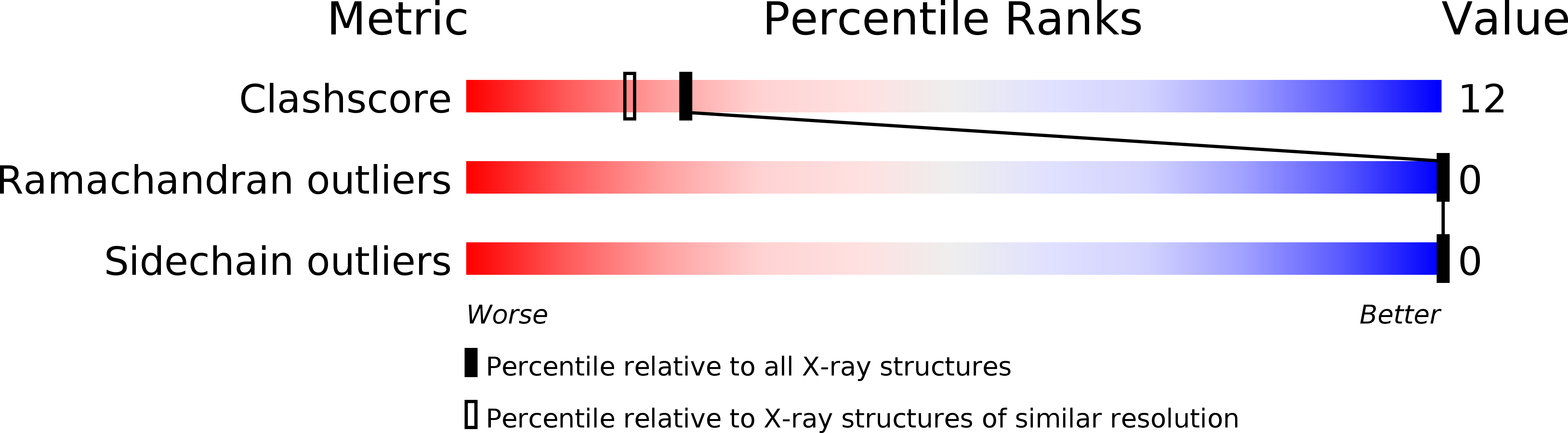
Crystal structure of rubredoxin from Desulfovibrio gigas to ultra-high 0.68 A resolution
Biological Source:
Source Organism:
Method Details:
Experimental Method:
Resolution:
0.68 Å
R-Value Free:
0.11
R-Value Work:
0.09
R-Value Observed:
0.09
Space Group:
P 1 21 1
Macromolecular Entities
Polymer Type:polypeptide(L)
Molecule:Rubredoxin
Chain IDs:A
Chain Length:52
Number of Molecules:1
Biological Source:Desulfovibrio gigas
Ligand Molecules
Primary Citation
Crystal structure of rubredoxin from Desulfovibrio gigas to ultra-high 0.68A resolution
Biochem.Biophys.Res.Commun. 349 79 90 (2006)
PMID: 16930541 DOI: 10.1016/j.bbrc.2006.07.205

Abstact

Rubredoxin (D.g. Rd) is a small non-heme iron-sulfur protein shown to function as a redox coupling protein from the sulfate reducing bacteria Desulfovibrio gigas. The protein is generally purified from anaerobic bacteria in which it is thought to be involved in electron transfer or exchange processes. Rd transfers an electron to oxygen to form water as part of a unique electron transfer chain, composed by NADH:rubredoxin oxidoreductase (NRO), rubredoxin and rubredoxin:oxygen oxidoreductase (ROO) in D.g. The crystal structure of D.g. Rd has been determined by means of both a Fe single-wavelength anomalous dispersion (SAD) signal and the direct method, and refined to an ultra-high 0.68 A resolution, using X-ray from a synchrotron. Rd contains one iron atom bound in a tetrahedral coordination by the sulfur atoms of four cysteinyl residues. Hydrophobic and pi-pi interactions maintain the internal Rd folding. Multiple conformations of the iron-sulfur cluster and amino acid residues are observed and indicate its unique mechanism of electron transfer. Several hydrogen bonds, including N-H...SG of the iron-sulfur, are revealed clearly in maps of electron density. Abundant waters bound to C-O peptides of residues Val8, Cys9, Gly10, Ala38, and Gly43, which may be involved in electron transfer. This ultrahigh-resolution structure allows us to study in great detail the relationship between structure and function of rubredoxin, such as salt bridges, hydrogen bonds, water structures, cysteine ligands, iron-sulfur cluster, and distributions of electron density among activity sites. For the first time, this information will provide a clear role for this protein in a strict anaerobic bacterium.

Legend

Protein

Chemical

Disease

Primary Citation of related structures