Deposition Date
2006-05-12
Release Date
2006-06-13
Last Version Date
2024-10-23
Entry Detail
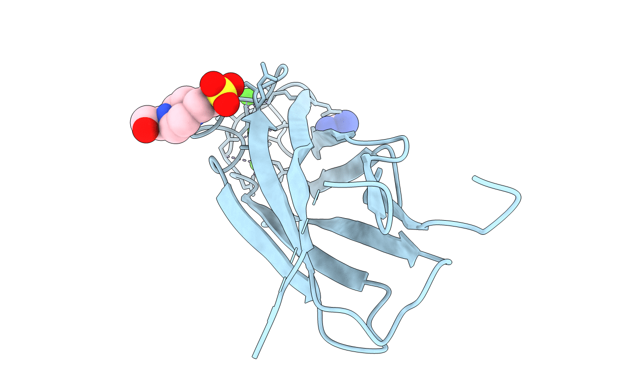
PDB ID:
2DPK
Keywords:
Title:
The Crystal Structure of the Primary Ca2+ Sensor of the Na+/Ca2+ Exchanger
Biological Source:
Source Organism(s):
Canis lupus familiaris (Taxon ID: 9615)
Expression System(s):
Method Details:
Experimental Method:
Resolution:
2.50 Å
R-Value Free:
0.28
R-Value Work:
0.22
R-Value Observed:
0.22
Space Group:
P 21 21 2