2DPE image
Deposition Date 2006-05-11
Release Date 2006-05-30
Last Version Date 2024-10-30
Entry Detail
PDB ID:
2DPE
Title:
Crystal structure of a secretory 40KDA glycoprotein from sheep at 2.0A resolution
Biological Source:
Source Organism:
Ovis aries (Taxon ID: 9940)
Method Details:
Experimental Method:
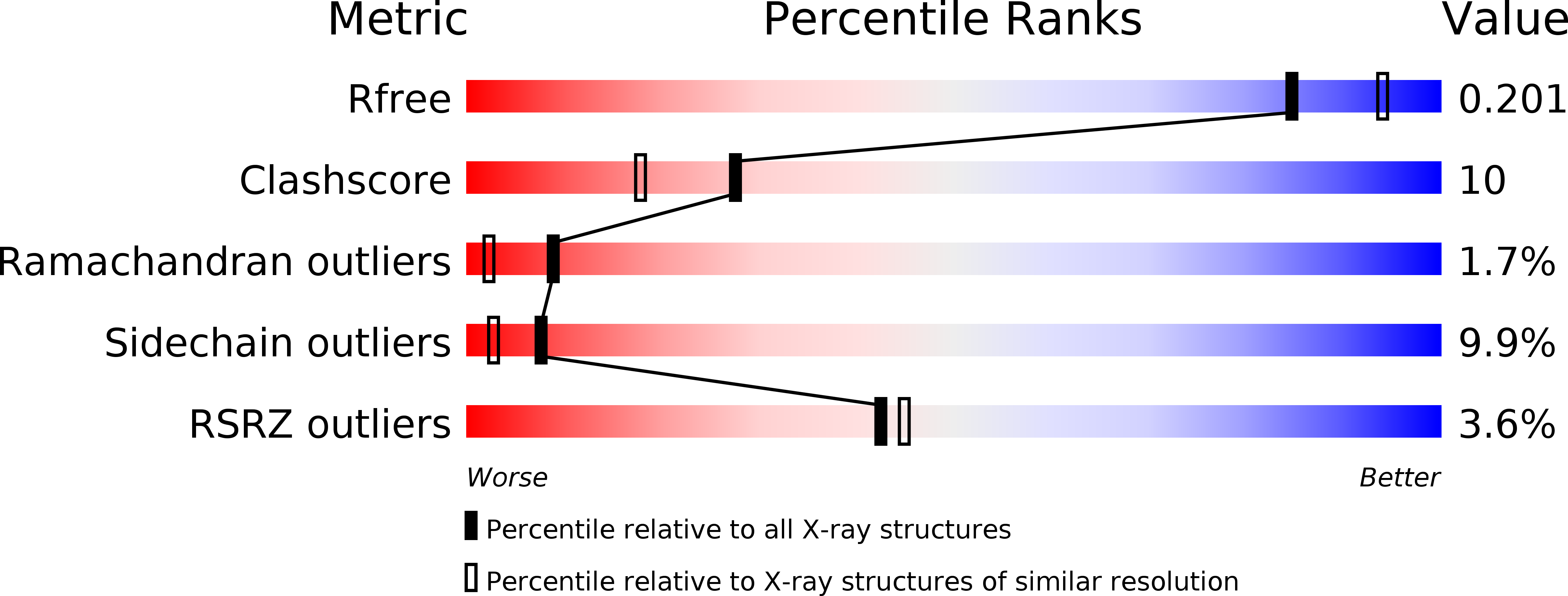
Resolution:
2.07 Å
R-Value Free:
0.20
R-Value Work:
0.18
R-Value Observed:
0.18
Space Group:
P 21 21 21
Macromolecular Entities
Polymer Type:polypeptide(L)
Molecule:Chitinase-3-like protein 1
Gene (Uniprot):CHI3L1
Chain IDs:A
Chain Length:361
Number of Molecules:1
Biological Source:Ovis aries
Modified Residue
Compound ID Chain ID Parent Comp ID Details 2D Image
ASN A ASN GLYCOSYLATION SITE
Primary Citation
Crystal structure of a secretory signalling glycoprotein from sheep at 2.0A resolution
J.Struct.Biol. 156 505 516 (2006)
PMID: 16859926 DOI: 10.1016/j.jsb.2006.05.008

Abstact

A 40kDa glycoprotein from dry secretion of sheep is implicated as a signaling factor and is named as SPS-40. This protein is secreted only during the early phase of involution when the drastic tissue remodeling occurs in the mammary gland. SPS-40 was purified from sheep dry secretions and crystallized using hanging drop vapour diffusion method. The crystals belong to orthorhombic space group P2(1)2(1)2(1) with cell dimensions, a=62.7A, b=66.4A, c=107.5A. The protein was also cloned for the determination of its complete amino acid sequence. The three-dimensional structure of SPS-40 was determined by X-ray crystallographic method at 2.0A resolution. The structure revealed the presence of an N-linked glycan chain at Asn39. The protein adopts a conformation with a classical (beta/alpha)(8)-barrel fold of triosephosphate isomerase (TIM) (residues 1-237 and 310-360) with an insertion of a small (alpha+beta) domain (residues 240-307) similar to that observed in chitinases. However, the Leu substitution for Glu in the consensus catalytic sequence in SPS-40 causes a loss of chitinase activity. Furthermore, the sugar-binding groove in SPS-40 is distorted considerably from the standard chitin-binding site in chitinase enzymes and hence the binding of chitin-like oligosaccharides is considerably hampered. Three surface loops, His188-His197, Phe202-Arg212 and Phe244-Pro260 have exceptionally high values of B-factors (average=70.5A(2)), indicating the presence of a less defined region.

Legend

Protein

Chemical

Disease

Primary Citation of related structures