2DOI image
Deposition Date 2006-04-29
Release Date 2006-12-05
Last Version Date 2024-10-16
Entry Detail
PDB ID:
2DOI
Keywords:
Title:
The X-ray crystallographic structure of the angiogenesis inhibitor, angiostatin, bound to a peptide from the group A streptococcus protein PAM
Biological Source:
Source Organism:
Homo sapiens (Taxon ID: 9606)
(Taxon ID: )
Host Organism:
Method Details:
Experimental Method:
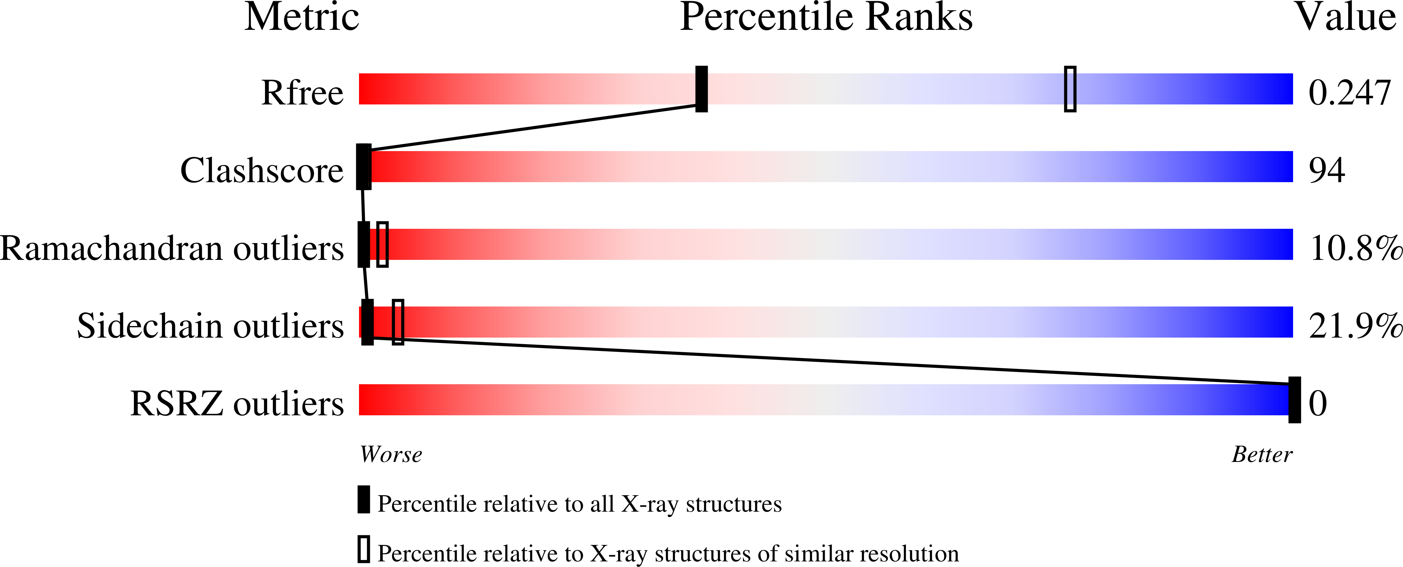
Resolution:
3.10 Å
R-Value Free:
0.29
R-Value Work:
0.20
R-Value Observed:
0.20
Space Group:
P 61
Macromolecular Entities
Polymer Type:polypeptide(L)
Molecule:Plasminogen-binding group A streptococcal M-like protein PAM
Gene (Uniprot):pam
Chain IDs:B (auth: C), D (auth: B)
Chain Length:30
Number of Molecules:2
Biological Source:
Polymer Type:polypeptide(L)
Molecule:Angiostatin
Gene (Uniprot):PLG
Mutations:N289E
Chain IDs:A (auth: X), C (auth: A)
Chain Length:234
Number of Molecules:2
Biological Source:Homo sapiens
Ligand Molecules
Primary Citation
X-ray crystallographic structure of the angiogenesis inhibitor, angiostatin, bound to a peptide from the group A streptococcal surface protein PAM
Biochemistry 45 11052 11060 (2006)
PMID: 16964966 DOI: 10.1021/bi060914j

Abstact

The crystal structure of the human Pg-derived angiogenesis inhibitor, angiostatin, complexed to VEK-30, a peptide from the group A streptococcal surface protein, PAM, was determined and refined to 2.3 A resolution. This is the first structure of angiostatin bound to a ligand and provides a model of the interaction between Pg and streptococcal-derived pathogenic proteins. VEK-30 contains a "through-space isostere" for C-terminal lysine, wherein Arg and Glu side chains, separated by one helical turn, bind within the bipolar angiostatin kringle 2 (K2) domain lysine-binding site. VEK-30 also makes several contacts with K2 residues that exist outside of the canonical LBS and are not conserved among the other Pg kringles, thus providing a molecular basis for the selectivity of VEK-30 for K2. The structure also shows that Pg kringle domains undergo significant structural rearrangement relative to one another and reveals dimerization between two molecules of angiostatin and VEK-30 related by crystallographic symmetry. This dimerization, which exists only in the crystal structure, is consistent with the parallel coiled-coil full-length PAM dimer expected from sequence similarities and homology modeling.

Legend

Protein

Chemical

Disease

Primary Citation of related structures