Deposition Date
1994-07-18
Release Date
1995-11-01
Last Version Date
2024-02-14
Entry Detail
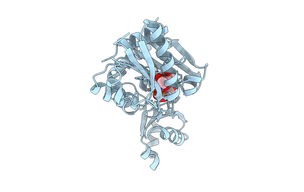
PDB ID:
2DLN
Keywords:
Title:
VANCOMYCIN RESISTANCE: STRUCTURE OF D-ALANINE:D-ALANINE LIGASE AT 2.3 ANGSTROMS RESOLUTION
Biological Source:
Source Organism(s):
Escherichia coli (Taxon ID: 562)
Method Details:
Experimental Method:
Resolution:
2.30 Å
R-Value Work:
0.17
R-Value Observed:
0.17
Space Group:
P 21 21 2