
Deposition Date
2006-04-11
Release Date
2006-10-24
Last Version Date
2024-03-13
Entry Detail
PDB ID:
2DKH
Keywords:
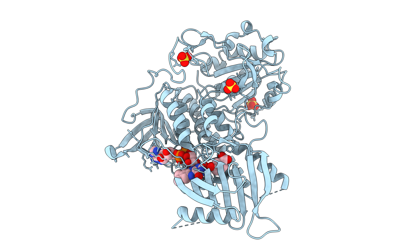
Title:
Crystal structure of 3-hydroxybenzoate hydroxylase from Comamonas testosteroni, in complex with the substrate
Biological Source:
Source Organism(s):
Comamonas testosteroni (Taxon ID: 285)
Method Details:
Experimental Method:
Resolution:
1.80 Å
R-Value Free:
0.19
R-Value Work:
0.17
R-Value Observed:
0.17
Space Group:
P 32 2 1
