
Deposition Date
2006-04-07
Release Date
2006-05-16
Last Version Date
2023-10-25
Entry Detail
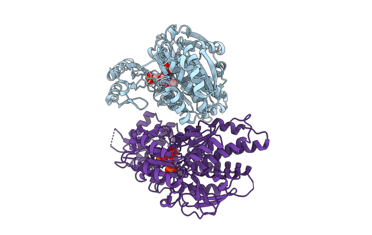
PDB ID:
2DKC
Keywords:
Title:
Crystal structure of N-acetylglucosamine-phosphate mutase, a member of the alpha-D-phosphohexomutase superfamily, in the substrate complex
Biological Source:
Source Organism(s):
Candida albicans (Taxon ID: 5476)
Expression System(s):
Method Details:
Experimental Method:
Resolution:
2.20 Å
R-Value Free:
0.24
R-Value Work:
0.18
R-Value Observed:
0.18
Space Group:
P 1 21 1
