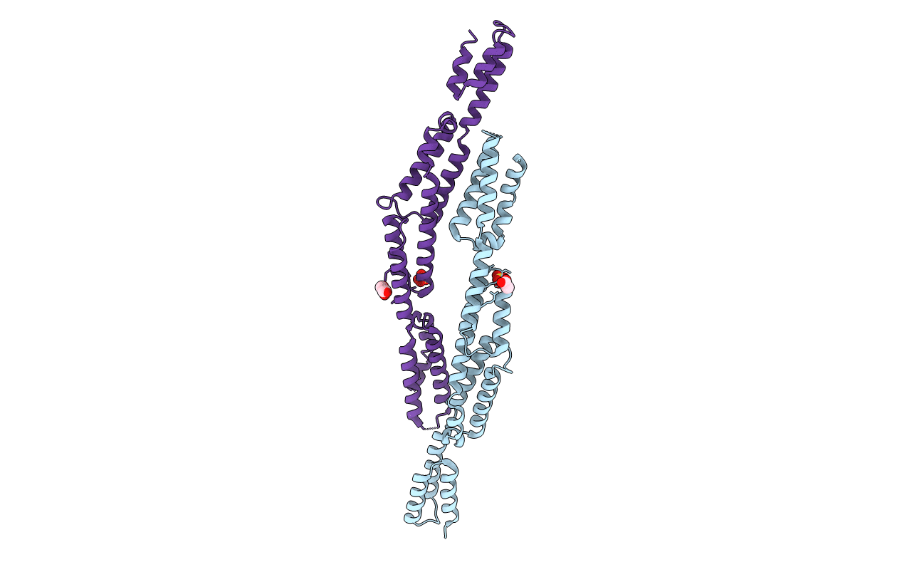
2DGJ image
Deposition Date 2006-03-14
Release Date 2007-03-20
Last Version Date 2024-10-23
Entry Detail
PDB ID:
2DGJ
Keywords:
Title:
Crystal structure of EbhA (756-1003 domain) from Staphylococcus aureus
Biological Source:
Source Organism:
Host Organism:
Method Details:
Experimental Method:
Resolution:
2.35 Å
R-Value Free:
0.28
R-Value Work:
0.24
R-Value Observed:
0.24
Space Group:
C 2 2 21
Macromolecular Entities
Polymer Type:polypeptide(L)
Molecule:hypothetical protein ebhA
Gene (Uniprot):ebhA
Chain IDs:A, B
Chain Length:257
Number of Molecules:2
Biological Source:Staphylococcus aureus
Modified Residue
Compound ID Chain ID Parent Comp ID Details 2D Image
MSE A MET SELENOMETHIONINE
Primary Citation
A helical string of alternately connected three-helix bundles for the cell wall-associated adhesion protein Ebh from Staphylococcus aureus
Structure 16 488 496 (2008)
PMID: 18334223 DOI: 10.1016/j.str.2007.12.018

Abstact

The 1.1 MDa cell-wall-associated adhesion protein of staphylococci, Ebh, consists of several distinct regions, including a large central region with 52 imperfect repeats of 126 amino acid residues. We investigated the structure of this giant molecule by X-ray crystallography, circular dichroism (CD) spectrometry, and small-angle X-ray scattering (SAXS). The crystal structure of two repeats showed that each repeat consists of two distinct three-helix bundles, and two such repeats are connected along the long axis, resulting in a rod-like structure that is 120 A in length. CD and SAXS analyses of the samples with longer repeats suggested that each repeat has an identical structure, and that such repeats are connected tandemly to form a rod-like structure in solution, the length of which increased proportionately with the number of repeating units. On the basis of these results, it was proposed that Ebh is a 320 nm rod-like molecule with some plasticity at module junctions.

Legend

Protein

Chemical

Disease

Primary Citation of related structures