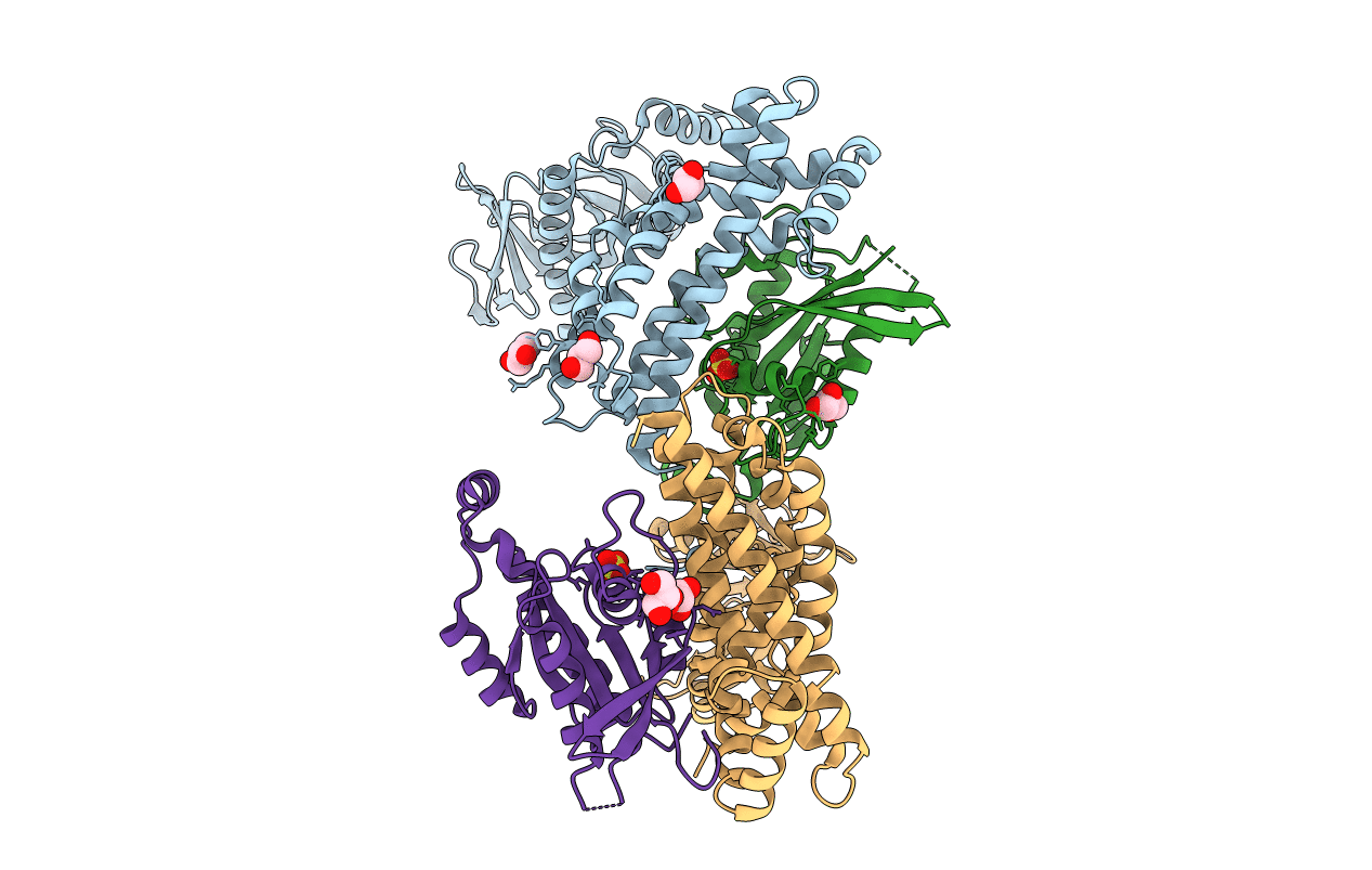
2DFK image
Deposition Date 2006-03-02
Release Date 2006-05-02
Last Version Date 2024-10-30
Entry Detail
PDB ID:
2DFK
Keywords:
Title:
Crystal structure of the CDC42-Collybistin II complex
Biological Source:
Source Organism:
Rattus norvegicus (Taxon ID: 10116)
Homo sapiens (Taxon ID: 9606)
Method Details:
Experimental Method:
Resolution:
2.15 Å
R-Value Free:
0.22
R-Value Work:
0.18
R-Value Observed:
0.18
Space Group:
P 21 21 21
Macromolecular Entities
Structures with similar UniProt ID
Protein Blast
Polymer Type:polypeptide(L)
Molecule:collybistin II
Gene (Uniprot):Arhgef9
Chain IDs:A, C
Chain Length:402
Number of Molecules:2
Biological Source:Rattus norvegicus
Structures with similar UniProt ID
Protein Blast
Polymer Type:polypeptide(L)
Molecule:cell division cycle 42 isoform 1
Gene (Uniprot):CDC42
Chain IDs:B, D
Chain Length:194
Number of Molecules:2
Biological Source:Homo sapiens
Primary Citation
The Crystal Structure of Cdc42 in Complex with Collybistin II, a Gephyrin-interacting Guanine Nucleotide Exchange Factor.
J.Mol.Biol. 359 35 46 (2006)
PMID: 16616186 DOI: 10.1016/j.jmb.2006.03.019

Abstact

The synaptic localization of ion channel receptors is essential for efficient synaptic transmission and the precise regulation of diverse neuronal functions. In the central nervous system, ion channel receptors reside in the postsynaptic membrane where they are juxtaposed to presynaptic terminals. For proper function, these ion channels have to be anchored to the cytoskeleton, and in the case of the inhibitory glycine and gamma-amino-butyric acid type A (GABA(A)) receptors this interaction is mediated by a gephyrin centered scaffold. Highlighting its central role in this receptor anchoring scaffold, gephyrin interacts with a number of proteins, including the neurospecific guanine nucleotide exchange factor collybistin. Collybistin belongs to the Dbl family of guanine nucleotide exchange factors, occurs in multiple splice variants, and is specific for Cdc42, a small GTPase belonging to the Rho family. The 2.3 Angstroms resolution crystal structure of the Cdc42-collybistin II complex reveals a novel conformation of the switch I region of Cdc42. It also provides the first direct observation of structural changes in the relative orientation of the Dbl-homology domain and the pleckstrin-homology domain in the same Dbl family protein. Biochemical data indicate that gephyrin negatively regulates collybistin activity.

Legend

Protein

Chemical

Disease

Primary Citation of related structures