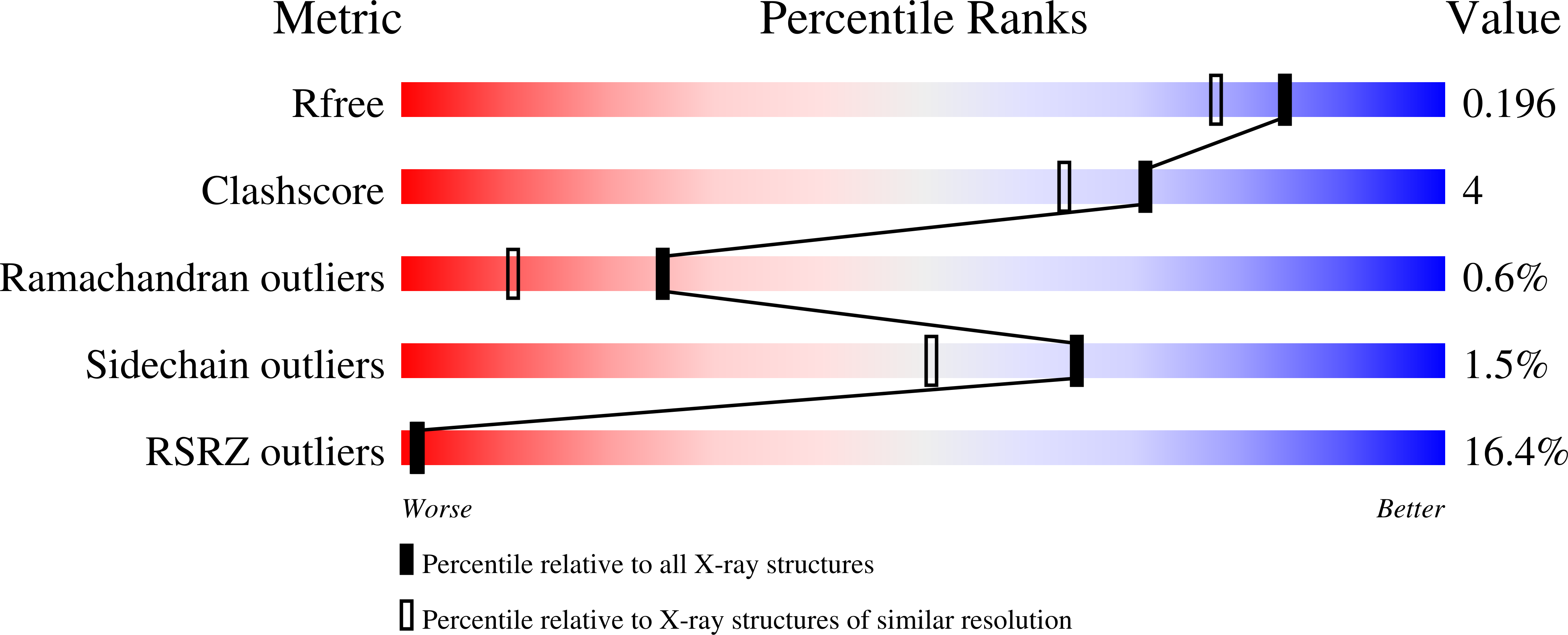
2DDF image
Deposition Date 2006-01-28
Release Date 2006-03-14
Last Version Date 2024-10-30
Entry Detail
PDB ID:
2DDF
Keywords:
Title:
Crystal structure of TACE in complex with TAPI-2
Biological Source:
Source Organism:
Homo sapiens (Taxon ID: 9606)
Host Organism:
Method Details:
Experimental Method:
Resolution:
1.70 Å
R-Value Free:
0.23
R-Value Work:
0.20
R-Value Observed:
0.20
Space Group:
P 21 21 21
Macromolecular Entities
Polymer Type:polypeptide(L)
Molecule:ADAM 17
Gene (Uniprot):ADAM17
Mutations:S266A, V353G, Q452N
Chain IDs:A, B
Chain Length:257
Number of Molecules:2
Biological Source:Homo sapiens
Peptide-like Molecules
PRD_000919
Primary Citation
Stabilization of the autoproteolysis of TNF-alpha converting enzyme (TACE) results in a novel crystal form suitable for structure-based drug design studies.
Protein Eng.Des.Sel. 19 155 161 (2006)
PMID: 16459338 DOI: 10.1093/protein/gzj014

Abstact

The crystallization of TNF-alpha converting enzyme (TACE) has been useful in understanding the structure-activity relationships of new chemical entities. However, the propensity of TACE to undergo autoproteolysis has made enzyme handling difficult and impeded the identification of inhibitor soakable crystal forms. The autoproteolysis of TACE was found to be specific (Y352-V353) and occurred within a flexible loop that is in close proximity to the P-side of the active site. The rate of autoproteolysis was found to be proportional to the concentration of TACE, suggesting a bimolecular reaction mechanism. A limited specificity study of the S(1)' subsite was conducted using surrogate peptides and suggested substitutions that would stabilize the proteolysis of the loop at positions Y352-V353. Two mutant proteases (V353G and V353S) were generated and proved to be highly resistant to autoproteolysis. The kinetics of the more resistant mutant (V353G) and wild-type TACE were compared and demonstrated virtually identical IC(50) values for a panel of competitive inhibitors. However, the k(cat)/K(m) of the mutant for a larger substrate (P6 - P(6)') was approximately 5-fold lower than that for the wild-type enzyme. Comparison of the complexed wild-type and mutant structures indicated a subtle shift in a peripheral P-side loop (comprising the mutation site) that may be involved in substrate binding/turnover and might explain the mild kinetic difference. The characterization of this stabilized form of TACE has yielded an enzyme with similar native kinetic properties and identified a novel crystal form that is suitable for inhibitor soaking and structure determination.

Legend

Protein

Chemical

Disease

Primary Citation of related structures