2DCU image
Deposition Date 2006-01-16
Release Date 2006-07-25
Last Version Date 2024-10-23
Entry Detail
PDB ID:
2DCU
Keywords:
Title:
Crystal structure of translation initiation factor aIF2betagamma heterodimer with GDP
Biological Source:
Source Organism:
Host Organism:
Method Details:
Experimental Method:
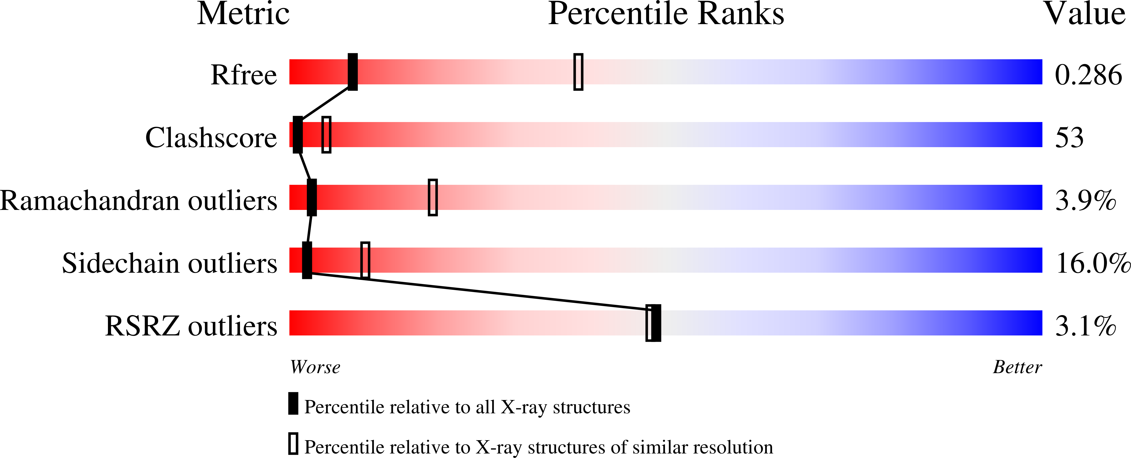
Resolution:
3.40 Å
R-Value Free:
0.29
R-Value Work:
0.24
Space Group:
P 21 21 21
Macromolecular Entities
Polymer Type:polypeptide(L)
Molecule:Translation initiation factor 2 gamma subunit
Gene (Uniprot):eif2g
Mutations:G236D
Chain IDs:A
Chain Length:419
Number of Molecules:1
Biological Source:Pyrococcus furiosus
Polymer Type:polypeptide(L)
Molecule:Translation initiation factor 2 beta subunit
Gene (Uniprot):eif2b
Chain IDs:B
Chain Length:148
Number of Molecules:1
Biological Source:Pyrococcus furiosus
Primary Citation
Structure of archaeal translational initiation factor 2 betagamma-GDP reveals significant conformational change of the beta-subunit and switch 1 region.
Proc.Natl.Acad.Sci.USA 103 13016 13021 (2006)
PMID: 16924118 DOI: 10.1073/pnas.0604165103

Abstact

Archaeal/eukaryotic initiation factor 2 (a/eIF2) consists of alpha-, beta-, and gamma-subunits and delivers initiator methionine tRNA (Met-tRNA(i)) to a small ribosomal subunit in a GTP-dependent manner. The structures of the aIF2betagamma (archaeal initiation factor 2 betagamma) heterodimeric complex in the apo and GDP forms were analyzed at 2.8- and 3.4-A resolution, respectively. The results showed that the N-terminal helix and the central helix-turn-helix domain of the beta-subunit bind to the G domain of the gamma-subunit but are distant from domains 2 and 3, to which the alpha-subunit and Met-tRNA(i) bind. This result is consistent with most of the previous analyses of eukaryotic factors, and thus indicates that the binding mode is essentially conserved among a/eIF2. Comparison with the uncomplexed structure showed significant differences between the two forms of the beta-subunit, particularly the C-terminal zinc-binding domain, which does not interact with the gamma-subunit and was suggested previously to be involved in GTP hydrolysis. Furthermore, the switch 1 region in the gamma-subunit, which is shown to be responsible for Met-tRNA(i) binding by mutational analysis, is moved away from the nucleotide through the interaction with highly conserved R87 in the beta-subunit. These results implicate that conformational change of the beta-subunit facilitates GTP hydrolysis by inducing the conformational change of the switch 1 region toward the off state.

Legend

Protein

Chemical

Disease

Primary Citation of related structures