
Deposition Date
2005-11-02
Release Date
2006-05-16
Last Version Date
2024-03-13
Entry Detail
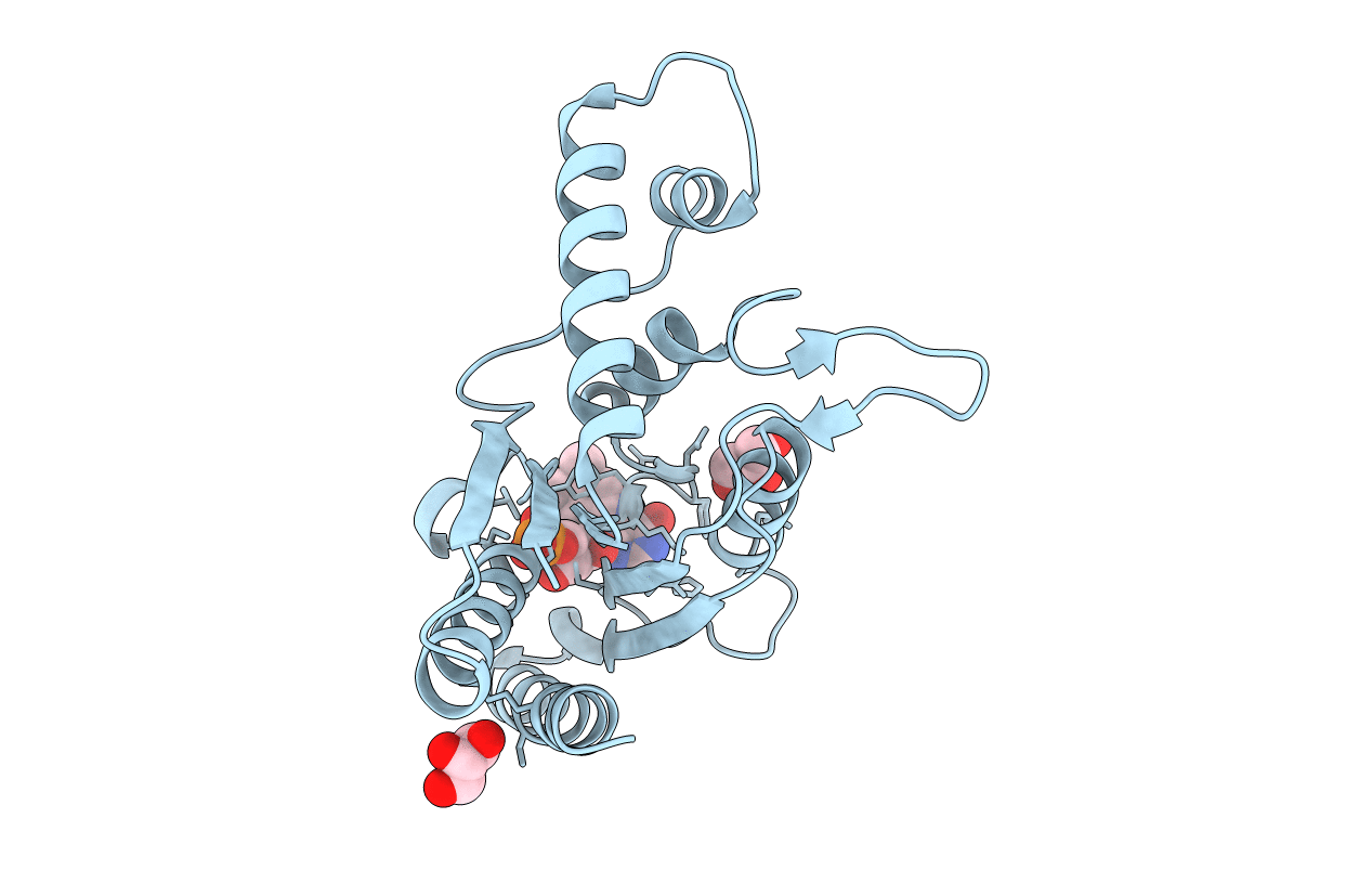
PDB ID:
2D5I
Keywords:
Title:
The crystal structure of AzoR (Azo Reductase) from Escherichia coli
Biological Source:
Source Organism(s):
Escherichia coli (Taxon ID: 83333)
Expression System(s):
Method Details:
Experimental Method:
Resolution:
2.20 Å
R-Value Free:
0.20
R-Value Work:
0.16
R-Value Observed:
0.17
Space Group:
P 4 21 2
