2D4A image
Deposition Date 2005-10-12
Release Date 2006-11-14
Last Version Date 2024-03-13
Entry Detail
PDB ID:
2D4A
Keywords:
Title:
Structure of the malate dehydrogenase from Aeropyrum pernix
Biological Source:
Source Organism:
Host Organism:
Method Details:
Experimental Method:
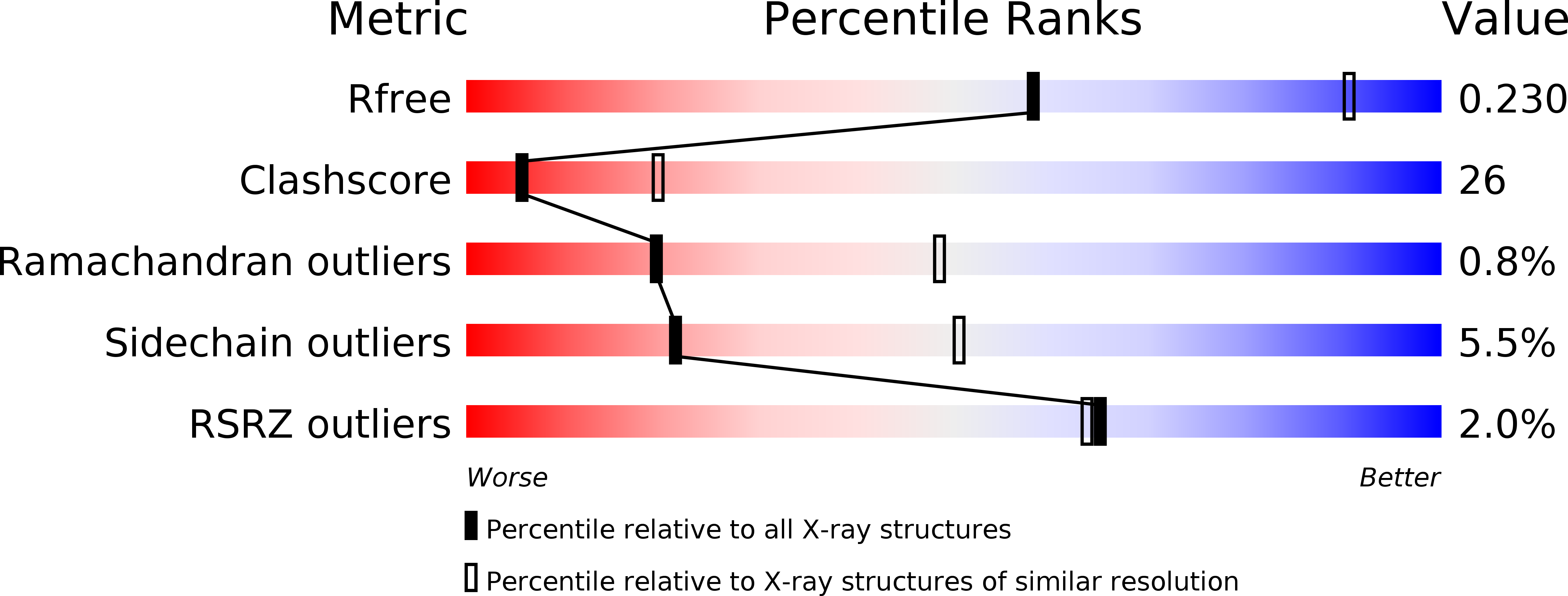
Resolution:
2.87 Å
R-Value Free:
0.24
R-Value Work:
0.20
R-Value Observed:
0.20
Space Group:
P 21 21 21
Macromolecular Entities
Polymer Type:polypeptide(L)
Molecule:Malate dehydrogenase
Gene (Uniprot):mdh
Chain IDs:A (auth: B), B (auth: D), C, D (auth: A)
Chain Length:308
Number of Molecules:4
Biological Source:Aeropyrum pernix
Primary Citation
Refolding, characterization and crystal structure of (S)-malate dehydrogenase from the hyperthermophilic archaeon Aeropyrum pernix.
Biochim.Biophys.Acta 1794 1496 1504 (2009)
PMID: 19555779 DOI: 10.1016/j.bbapap.2009.06.014

Abstact

Tartrate oxidation activity was found in the crude extract of an aerobic hyperthermophilic archaeon Aeropyrum pernix, and the enzyme was identified as (S)-malate dehydrogenase (MDH), which, when produced in Escherichia coli, was mainly obtained as an inactive inclusion body. The inclusion body was dissolved in 6 M guanidine-HCl and gradually refolded to the active enzyme through dilution of the denaturant. The purified recombinant enzyme consisted of four identical subunits with a molecular mass of about 110 kDa. NADP was preferred as a coenzyme over NAD for (S)-malate oxidation and, unlike MDHs from other sources, this enzyme readily catalyzed the oxidation of (2S,3S)-tartrate and (2S,3R)-tartrate. The tartrate oxidation activity was also observed in MDHs from the hyperthermophilic archaea Methanocaldococcus jannaschii and Archaeoglobus fulgidus, suggesting these hyperthermophilic MDHs loosely bind their substrates. The refolded A. pernix MDH was also crystallized, and the structure was determined at a resolution of 2.9 A. Its overall structure was similar to those of the M. jannaschii, Chloroflexus aurantiacus, Chlorobium vibrioforme and Cryptosporidium parvum [lactate dehydrogenase-like] MDHs with root-mean-square-deviation values between 1.4 and 2.1 A. Consistent with earlier reports, Ala at position 53 was responsible for coenzyme specificity, and the next residue, Arg, was important for NADP binding. Structural comparison revealed that the hyperthermostability of the A. pernix MDH is likely attributable to its smaller cavity volume and larger numbers of ion pairs and ion-pair networks, but the molecular strategy for thermostability may be specific for each enzyme.

Legend

Protein

Chemical

Disease

Primary Citation of related structures