2D3Y image
Deposition Date 2005-10-04
Release Date 2006-10-17
Last Version Date 2024-03-13
Entry Detail
PDB ID:
2D3Y
Keywords:
Title:
Crystal structure of uracil-DNA glycosylase from Thermus Thermophilus HB8
Biological Source:
Source Organism:
Method Details:
Experimental Method:
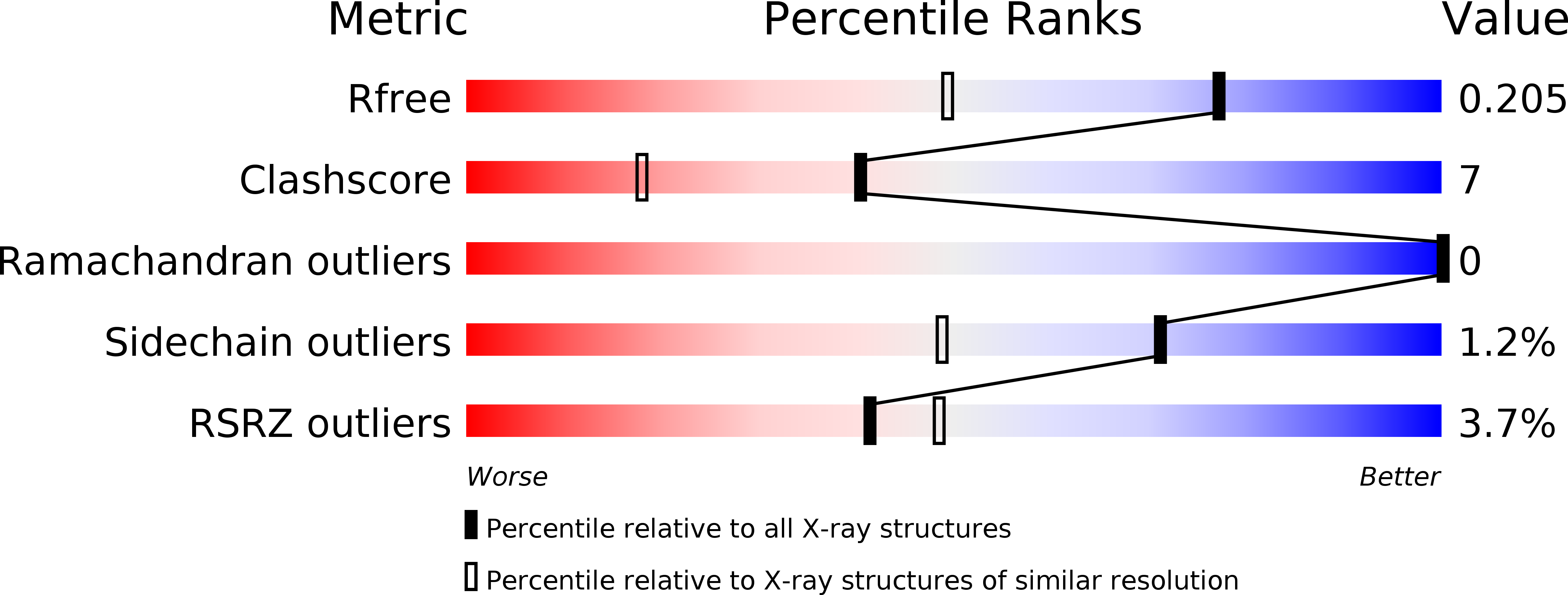
Resolution:
1.55 Å
R-Value Free:
0.21
R-Value Work:
0.18
Space Group:
P 21 21 21
Macromolecular Entities
Polymer Type:polypeptide(L)
Molecule:uracil-DNA glycosylase
Gene (Uniprot):udgb
Chain IDs:A
Chain Length:219
Number of Molecules:1
Biological Source:Thermus thermophilus
Primary Citation
Crystal structure of family 5 uracil-DNA glycosylase bound to DNA.
J.Mol.Biol. 373 839 850 (2007)
PMID: 17870091 DOI: 10.1016/j.jmb.2007.08.022

Abstact

Uracil-DNA glycosylase (UDG) removes uracil generated by the deamination of cytosine or misincorporation of deoxyuridine monophosphate. Within the UDG superfamily, a fifth UDG family lacks a polar residue in the active-site motif, which mediates the hydrolysis of the glycosidic bond by activation of a water molecule in UDG families 1-4. We have determined the crystal structure of a novel family 5 UDG from Thermus thermophilus HB8 complexed with DNA containing an abasic site. The active-site structure suggests this enzyme uses both steric force and water activation for its excision reaction. A conserved asparagine residue acts as a ligand to the catalytic water molecule. The structure also implies that another water molecule acts as a barrier during substrate recognition. Based on no significant open-closed conformational change upon binding to DNA, we propose a "slide-in" mechanism for initial damage recognition.

Legend

Protein

Chemical

Disease

Primary Citation of related structures