
Deposition Date
2005-10-03
Release Date
2006-01-17
Last Version Date
2024-03-13
Entry Detail
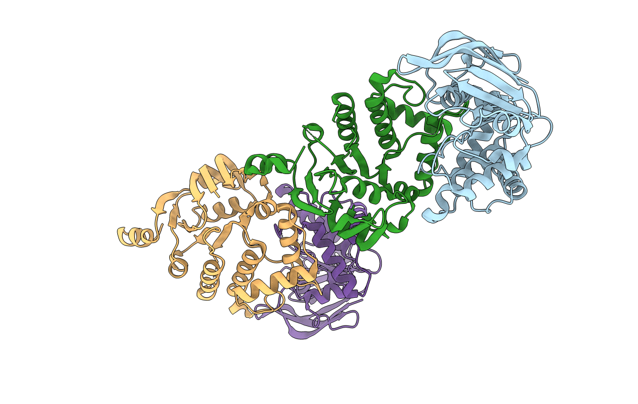
PDB ID:
2D3W
Keywords:
Title:
Crystal Structure of Escherichia coli SufC, an ATPase compenent of the SUF iron-sulfur cluster assembly machinery
Biological Source:
Source Organism(s):
Escherichia coli (Taxon ID: 562)
Expression System(s):
Method Details:
Experimental Method:
Resolution:
2.50 Å
R-Value Free:
0.29
R-Value Work:
0.22
R-Value Observed:
0.23
Space Group:
P 1 21 1
