2D3K image
Deposition Date 2005-09-29
Release Date 2006-03-29
Last Version Date 2023-10-25
Entry Detail
PDB ID:
2D3K
Keywords:
Title:
Structural study on Project ID PH1539 from Pyrococcus horikoshii OT3
Biological Source:
Source Organism:
Host Organism:
Method Details:
Experimental Method:
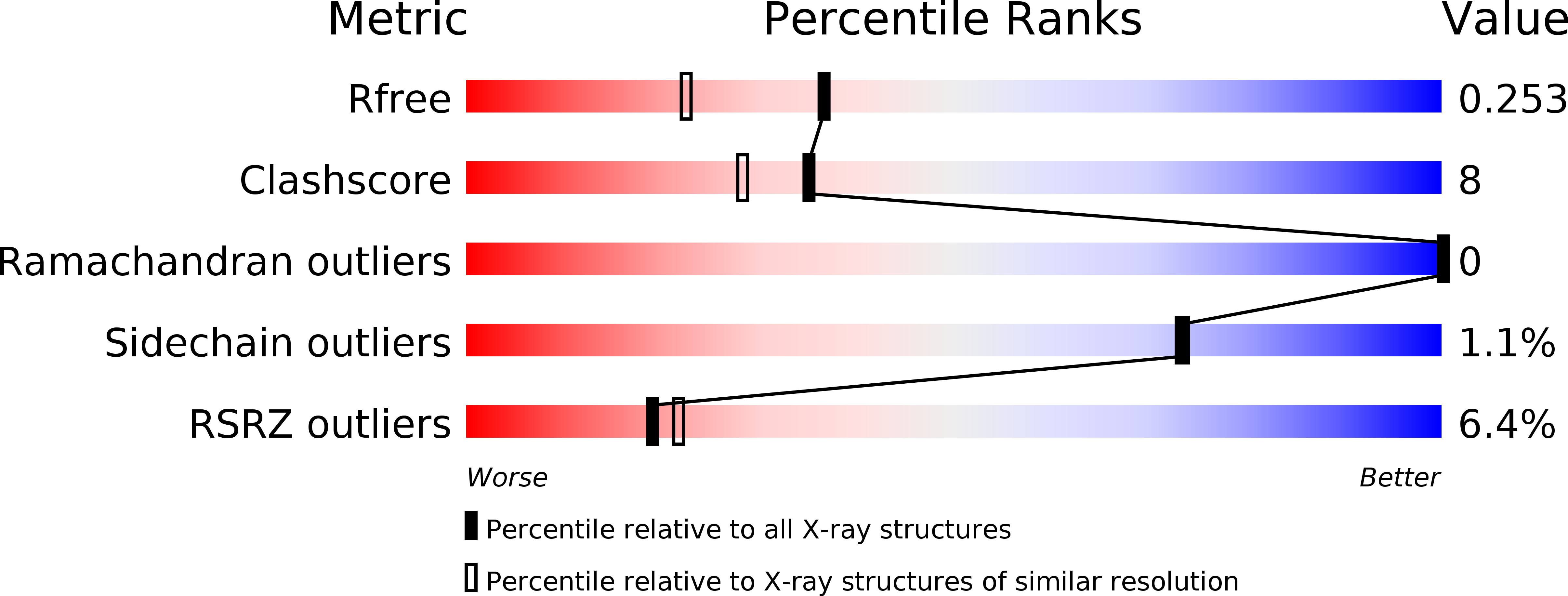
Resolution:
1.90 Å
R-Value Free:
0.25
R-Value Work:
0.20
R-Value Observed:
0.20
Space Group:
P 43 2 2
Macromolecular Entities
Polymer Type:polypeptide(L)
Molecule:Peptidyl-tRNA hydrolase
Gene (Uniprot):pth
Chain IDs:A, B
Chain Length:121
Number of Molecules:2
Biological Source:Pyrococcus horikoshii
Ligand Molecules
Primary Citation
Structure of peptidyl-tRNA hydrolase 2 from Pyrococcus horikoshii OT3: insight into the functional role of its dimeric state.
Acta Crystallogr.,Sect.D 64 444 453 (2008)
PMID: 18391411 DOI: 10.1107/S0907444908002850

Abstact

Peptidyl-tRNA hydrolases catalyze the hydrolytic removal of the peptidyl moiety from the peptidyl-tRNA molecule to prevent misreading during translation. Here, the expression, purification, crystallization and X-ray diffraction study of peptidyl-tRNA hydrolase 2 from Pyrococcus horikoshii OT3 (PhPth2) are described. The crystal structures were determined as similar biological dimers in two different forms: P4(1)2(1)2 at 1.2 A resolution (form 1) and P4(3)22 at 1.9 A resolution (form 2). In the form 1 structure, the asymmetric unit contains one PhPth2 subunit and a crystallographic twofold axis defines the dimeric association with the cognate subunit. In the form 2 structure, there are two PhPth2 subunits in the asymmetric unit that make a similar dimer with a noncrystallographic twofold axis. In order to evaluate the thermodynamic stability, the intra-protomer and inter-protomer interactions of PhPth2 were analyzed and compared with those of other Pth2-family members. The thermodynamic parameters show that the large number of ion pairs compared with family members from other mesophilic organisms would contribute to the thermostability of PhPth2. The structural difference between the two dimers was quantitatively evaluated by a multiple C(alpha)-atom superposition. A significant structural difference between the two dimers was observed around the putative active site of this enzyme. A rigid-body rotation takes place so as to retain the dimeric twofold symmetry, suggesting positive cooperativity upon tRNA binding. The mechanism of ligand binding was further investigated using a docking model with a tRNA molecule. The docking study suggests that the binding of tRNA requires its simultaneous interaction with both subunits of the PhPth2 dimer.

Legend

Protein

Chemical

Disease

Primary Citation of related structures