2D3I image
Deposition Date 2005-09-28
Release Date 2005-11-29
Last Version Date 2024-11-13
Entry Detail
PDB ID:
2D3I
Keywords:
Title:
Crystal Structure of Aluminum-Bound Ovotransferrin at 2.15 Angstrom Resolution
Biological Source:
Source Organism:
Gallus gallus (Taxon ID: 9031)
Method Details:
Experimental Method:
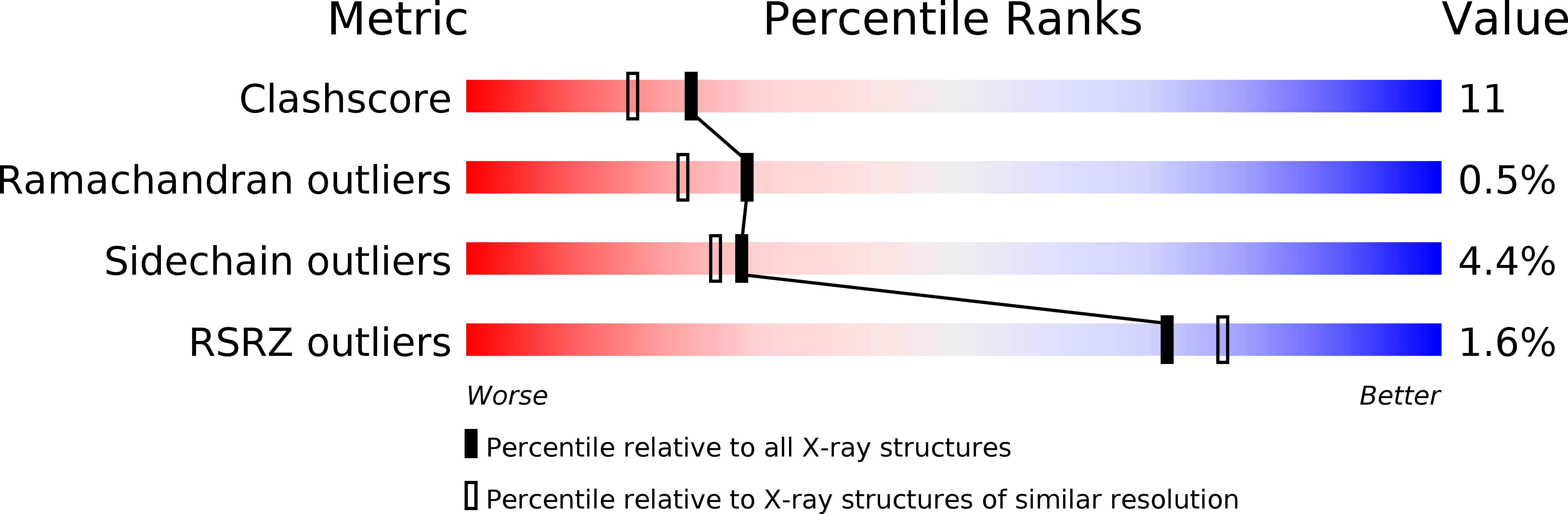
Resolution:
2.15 Å
R-Value Free:
0.24
R-Value Work:
0.20
R-Value Observed:
0.20
Space Group:
P 1 21 1
Macromolecular Entities
Structures with similar UniProt ID
Protein Blast
Polymer Type:polypeptide(L)
Molecule:Ovotransferrin
Chain IDs:A
Chain Length:686
Number of Molecules:1
Biological Source:Gallus gallus
Primary Citation
Structure of aluminium-bound ovotransferrin at 2.15 Angstroms resolution.
Acta Crystallogr.,Sect.D 61 1636 1642 (2005)
PMID: 16301797 DOI: 10.1107/S090744490503266X

Abstact

Transferrin, well known as an iron-transport protein, can bind other metal ions, including toxic ones, and is considered to play an important role in the transportation of such metal ions. Here, a crystal structure of aluminium-bound transferrin is described for the first time. Colourless needle-shaped crystals of aluminium-bound ovotransferrin were obtained in PEG 400 solution. Structural determination was performed by molecular replacement using diferric (iron-bound) ovotransferrin as a model and the structural refinement was performed in the 50-2.15 Angstroms resolution range. The overall organization of the aluminium-bound form is almost the same as the iron-bound form: the protein is folded into two homologous lobes (N- and C-lobes) with two domains; two metal-binding sites are located within the inter-domain clefts of each lobe. Four residues (one Asp, two Tyr and one His) and one bicarbonate anion were found to bind an aluminium ion in either lobe, as in the iron-bound form. The highly similar domain-closed structure of the Al(3+)-bound form may permit the binding of Al(3+)-bound transferrin to the transferrin receptor. An unusual interaction, the dilysine trigger, which facilitates iron release at low pH in the endosome, was also found in the Al(3+)-bound form. These findings support the participation of transferrin in the transport of Al(3+) ions in vivo.

Legend

Protein

Chemical

Disease

Primary Citation of related structures
Feedback Form
Name
Email
Institute
Feedback