Deposition Date
2005-09-27
Release Date
2006-02-14
Last Version Date
2024-03-13
Entry Detail
PDB ID:
2D3D
Keywords:
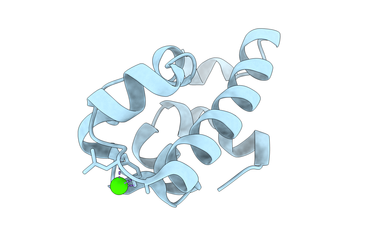
Title:
crystal structure of the RNA binding SAM domain of saccharomyces cerevisiae Vts1
Biological Source:
Source Organism(s):
Saccharomyces cerevisiae (Taxon ID: 4932)
Expression System(s):
Method Details:
Experimental Method:
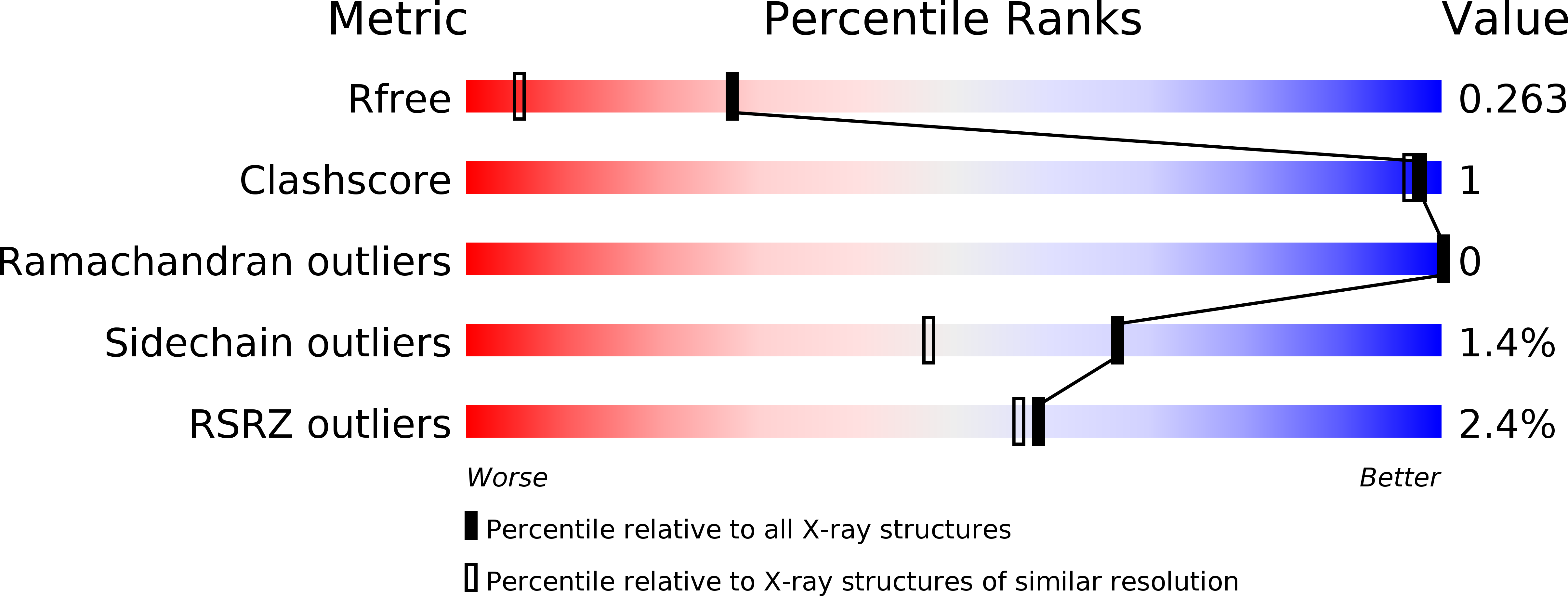
Resolution:
1.60 Å
R-Value Free:
0.25
R-Value Work:
0.20
R-Value Observed:
0.20
Space Group:
P 21 21 21