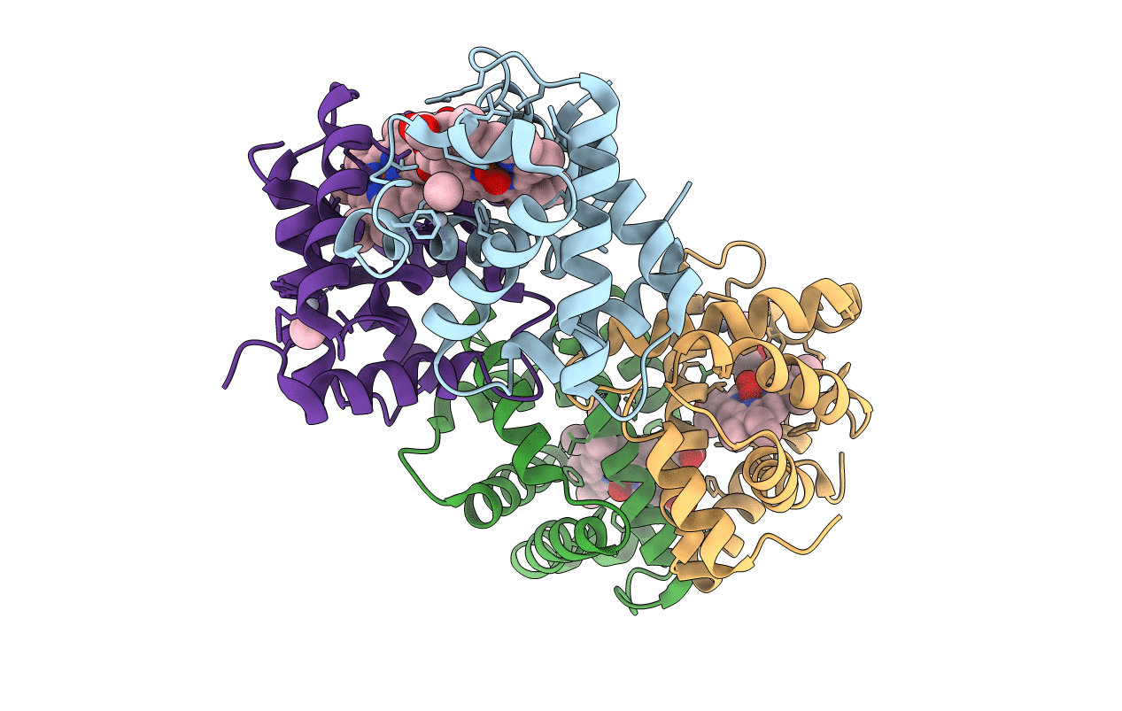
2D2N image
Entry Detail
PDB ID:
2D2N
Title:
Structure of an extracellular giant hemoglobin of the gutless beard worm Oligobrachia mashikoi
Biological Source:
Source Organism:
PDB Version:
Deposition Date:
2005-09-12
Release Date:
2005-10-25
Method Details:
Experimental Method:
Resolution:
3.20 Å
R-Value Free:
0.29
R-Value Work:
0.24
Space Group:
H 3 2
Macromolecular Entities
Polymer Type:polypeptide(L)
Description:Giant hemoglobin, A1(b) globin chain
Chain IDs:A
Chain Length:140
Number of Molecules:1
Biological Source:Oligobrachia mashikoi
Polymer Type:polypeptide(L)
Description:Giant hemoglobin, A2(a5) globin chain
Chain IDs:B
Chain Length:142
Number of Molecules:1
Biological Source:Oligobrachia mashikoi
Polymer Type:polypeptide(L)
Description:Giant hemoglobin, B2(c) globin chain
Chain IDs:C
Chain Length:147
Number of Molecules:1
Biological Source:Oligobrachia mashikoi
Polymer Type:polypeptide(L)
Description:Giant hemoglobin, B1(d) globin chain
Chain IDs:D
Chain Length:145
Number of Molecules:1
Biological Source:Oligobrachia mashikoi
Primary Citation
Structure of an extracellular giant hemoglobin of the gutless beard worm Oligobrachia mashikoi.
Proc.Natl.Acad.Sci.USA 102 14521 14526 (2005)
PMID: 16204001 DOI: 10.1073/pnas.0501541102

Abstact

Mouthless and gutless marine animals, pogonophorans and vestimentiferans, obtain their nutrition solely from their symbiotic chemoautotrophic sulfur-oxidizing bacteria. These animals have sulfide-binding 400-kDa and/or 3,500-kDa Hb, which transports oxygen and sulfide simultaneously. The symbiotic bacteria are supplied with sulfide by Hb of the host animal and use it to provide carbon compounds. Here, we report the crystal structure of a 400-kDa Hb from pogonophoran Oligobrachia mashikoi at 2.85-A resolution. The structure is hollow-spherical, composed of a total of 24 globins as a dimer of dodecamer. This dodecameric assemblage would be a fundamental structural unit of both 400-kDa and 3,500-kDa Hbs. The structure of the mercury derivative used for phasing provides insights into the sulfide-binding mechanism. The mercury compounds bound to all free Cys residues that have been expected as sulfide-binding sites. Some of the free Cys residues are surrounded by Phe aromatic rings, and mercury atoms come into contact with these residues in the derivative structure. It is strongly suggested that sulfur atoms bound to these sites could be stabilized by aromatic-electrostatic interactions by the surrounding Phe residues.

Legend

Protein

Chemical

Disease

Primary Citation of related structures