2D1V image
Deposition Date 2005-09-01
Release Date 2006-09-12
Last Version Date 2024-03-13
Entry Detail
PDB ID:
2D1V
Keywords:
Title:
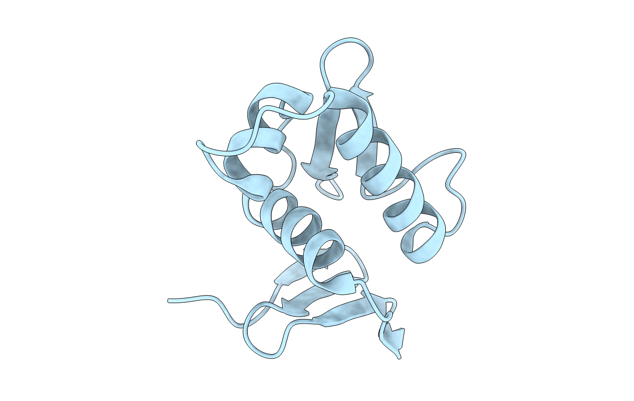
Crystal structure of DNA-binding domain of Bacillus subtilis YycF
Biological Source:
Source Organism:
Host Organism:
Method Details:
Experimental Method:
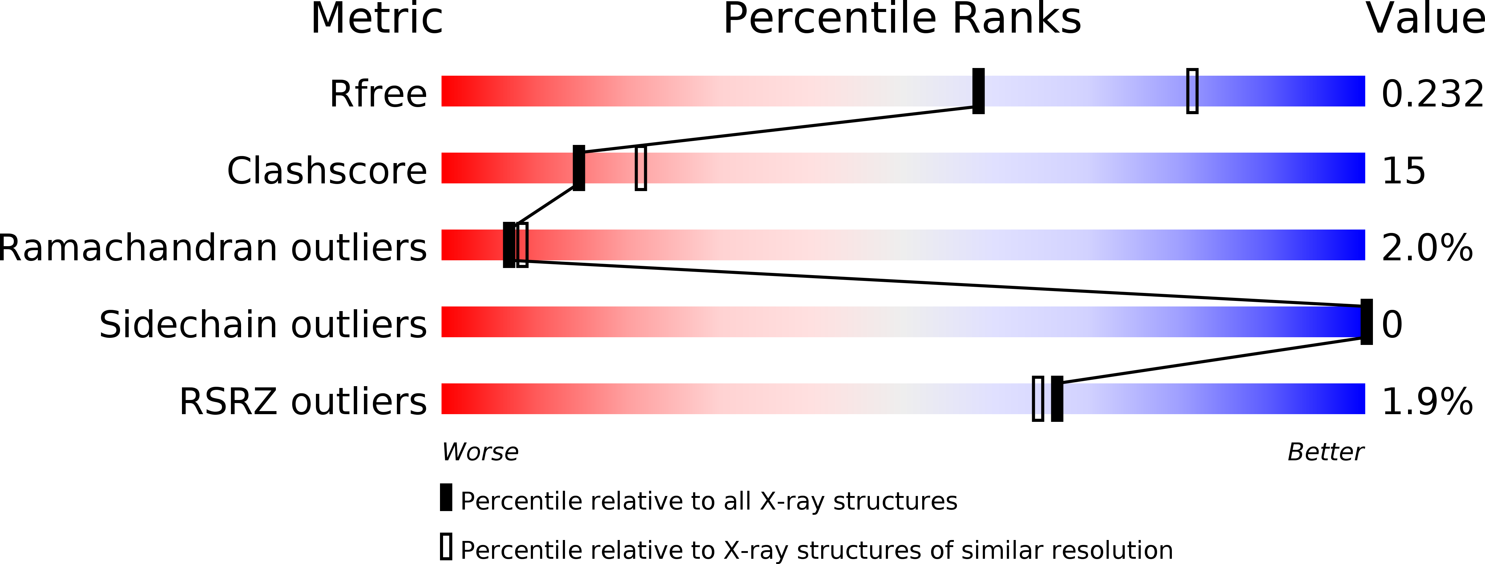
Resolution:
2.40 Å
R-Value Free:
0.23
R-Value Work:
0.22
R-Value Observed:
0.22
Space Group:
C 2 2 21
Macromolecular Entities
Structures with similar UniProt ID
Protein Blast
Polymer Type:polypeptide(L)
Molecule:Transcriptional regulatory protein yycF
Gene (Uniprot):walR
Chain IDs:A
Chain Length:108
Number of Molecules:1
Biological Source:Bacillus subtilis
Primary Citation
Response regulator YycF essential for bacterial growth: X-ray crystal structure of the DNA-binding domain and its PhoB-like DNA recognition motif
Febs Lett. 582 3434 3438 (2008)
PMID: 18789936 DOI: 10.1016/j.febslet.2008.09.007

Abstact

A response regulator YycF and its cognate sensor kinase YycG constitute the two-component signal transduction system essential for growth of Gram-positive bacteria with a low GC content. We have determined the X-ray crystal structure of the effector domain of Bacillus subtilis YycF involved in DNA binding. The structure, containing a winged helix-turn-helix motif, was found to be very similar to that of the response regulator PhoB from Escherichia coli. Specific binding of YycF to the PhoB-regulated alkaline phosphatase promoter was also demonstrated.

Legend

Protein

Chemical

Disease

Primary Citation of related structures
Feedback Form
Name
Email
Institute
Feedback