Deposition Date
2005-08-27
Release Date
2006-09-12
Last Version Date
2024-10-30
Entry Detail
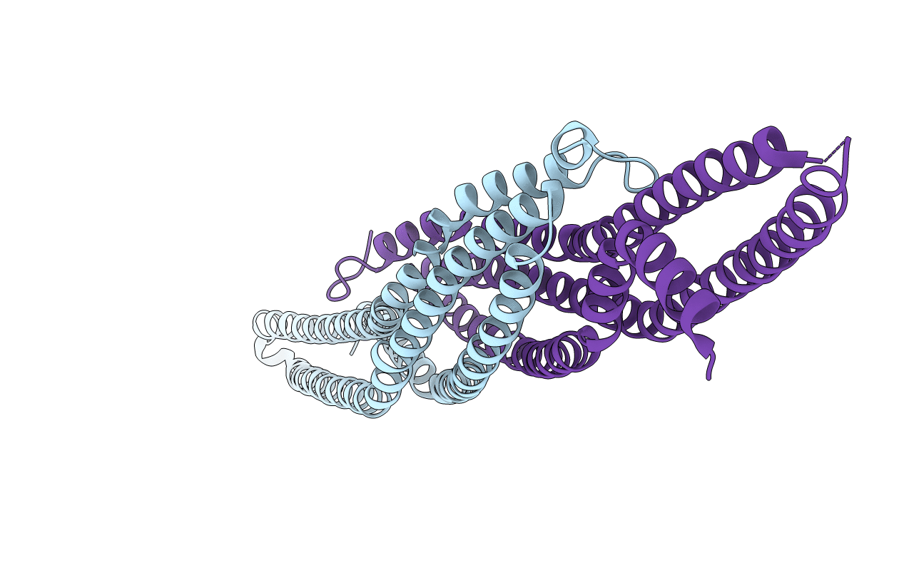
PDB ID:
2D1L
Keywords:
Title:
Structure of F-actin binding domain IMD of MIM (Missing In Metastasis)
Biological Source:
Source Organism(s):
Mus musculus (Taxon ID: 10090)
Expression System(s):
Method Details:
Experimental Method:
Resolution:
1.85 Å
R-Value Free:
0.22
R-Value Work:
0.18
R-Value Observed:
0.18
Space Group:
P 1 21 1