2D11 image
Deposition Date 2005-08-11
Release Date 2006-07-18
Last Version Date 2024-03-13
Entry Detail
PDB ID:
2D11
Keywords:
Title:
Crystal structure of the Radixin FERM domain complexed with the NHERF-2 C-terminal tail peptide
Biological Source:
Source Organism:
Mus musculus (Taxon ID: 10090)
(Taxon ID: )
Host Organism:
Method Details:
Experimental Method:
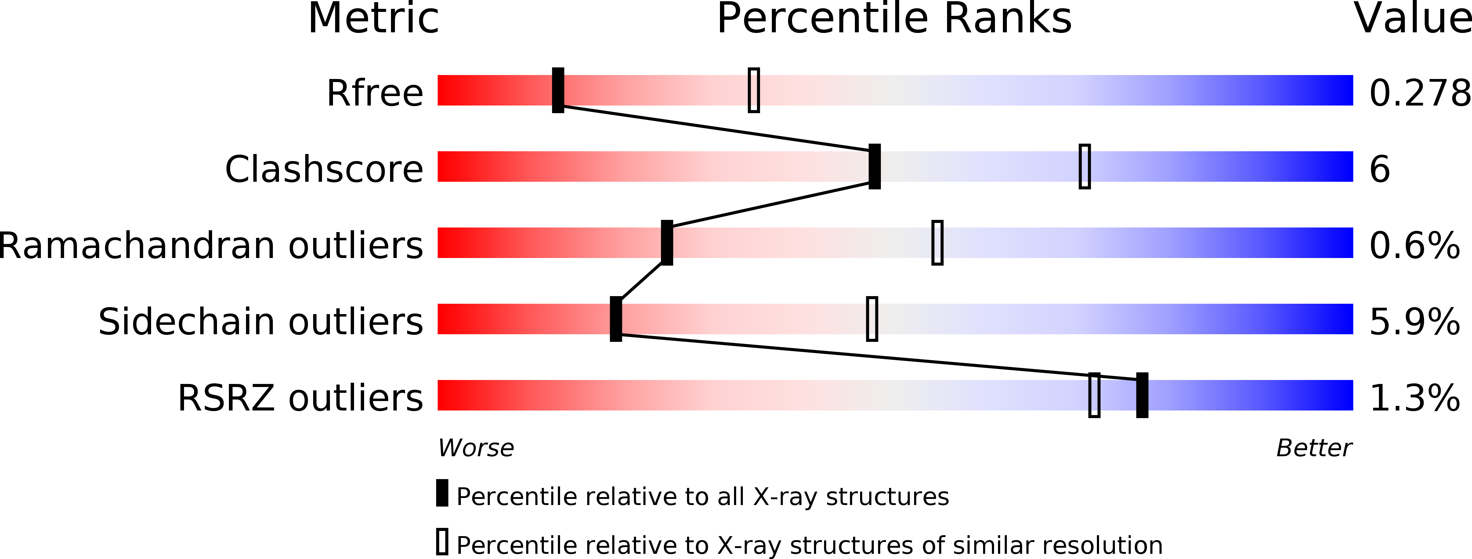
Resolution:
2.81 Å
R-Value Free:
0.27
R-Value Work:
0.22
R-Value Observed:
0.22
Space Group:
P 21 21 21
Macromolecular Entities
Polymer Type:polypeptide(L)
Molecule:Radixin
Gene (Uniprot):Rdx
Chain IDs:A, B, C, D
Chain Length:312
Number of Molecules:4
Biological Source:Mus musculus
Polymer Type:polypeptide(L)
Molecule:Na(+)/H(+) exchange regulatory cofactor NHE-RF2
Gene (Uniprot):NHERF2
Chain IDs:E, F, G, H
Chain Length:28
Number of Molecules:4
Biological Source:
Primary Citation

Abstact

The Na+/H+ exchanger regulatory factor (NHERF) is a key adaptor protein involved in the anchoring of ion channels and receptors to the actin cytoskeleton through binding to ERM (ezrin/radixin/moesin) proteins. NHERF binds the FERM domain of ERM proteins, although NHERF has no signature Motif-1 sequence for FERM binding found in adhesion molecules. The crystal structures of the radixin FERM domain complexed with the NHERF-1 and NHERF-2 C-terminal peptides revealed a peptide binding site of the FERM domain specific for the 13 residue motif MDWxxxxx(L/I)Fxx(L/F) (Motif-2), which is distinct from Motif-1. This Motif-2 forms an amphipathic alpha helix for hydrophobic docking to subdomain C of the FERM domain. This docking causes induced-fit conformational changes in subdomain C and affects binding to adhesion molecule peptides, while the two binding sites are not overlapped. Our studies provide structural paradigms for versatile ERM linkages between membrane proteins and the cytoskeleton.

Legend

Protein

Chemical

Disease

Primary Citation of related structures