2CWF image
Deposition Date 2005-06-20
Release Date 2005-10-04
Last Version Date 2024-03-13
Entry Detail
PDB ID:
2CWF
Keywords:
Title:
Crystal Structure of delta1-piperideine-2-carboxylate reductase from Pseudomonas syringae complexed with NADPH
Biological Source:
Source Organism:
Method Details:
Experimental Method:
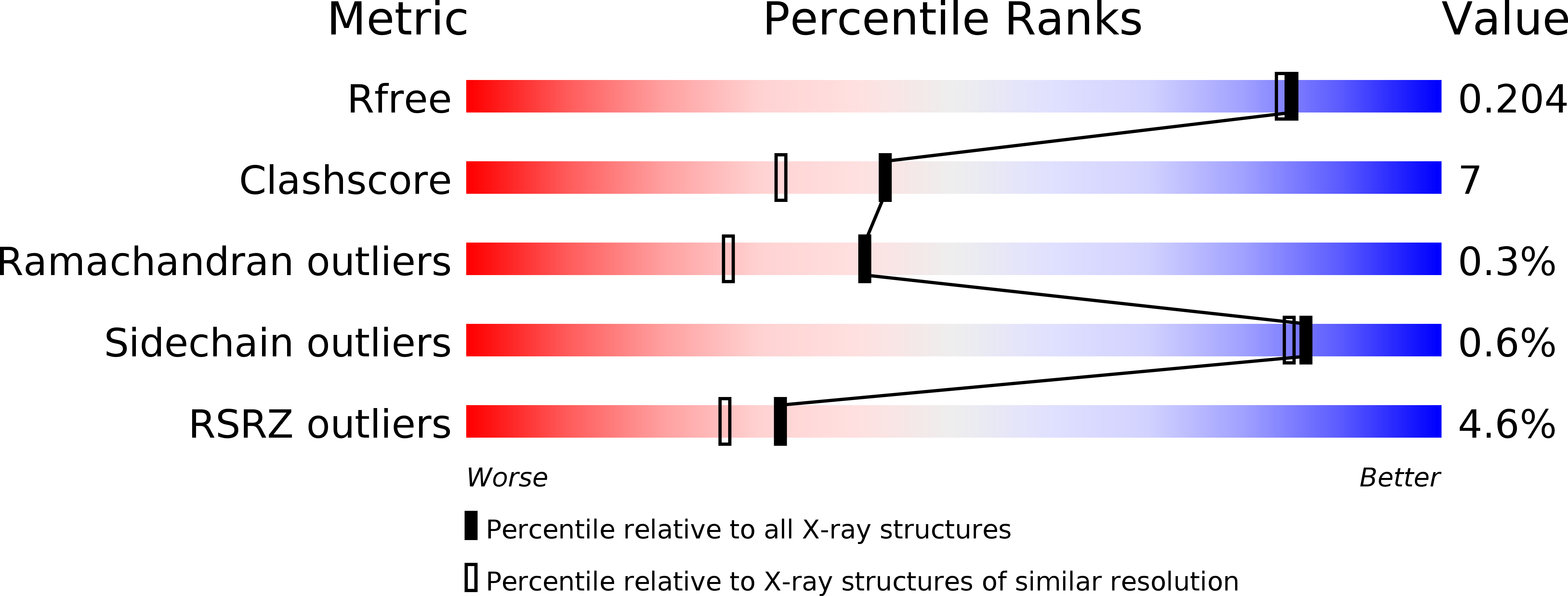
Resolution:
1.80 Å
R-Value Free:
0.20
R-Value Work:
0.18
R-Value Observed:
0.18
Space Group:
P 21 21 21
Macromolecular Entities
Polymer Type:polypeptide(L)
Molecule:delta1-piperideine-2-carboxylate reductase
Gene (Uniprot):dpkA
Chain IDs:A, B
Chain Length:343
Number of Molecules:2
Biological Source:Pseudomonas syringae pv. tomato
Ligand Molecules
Primary Citation
Crystal structures of Delta1-piperideine-2-carboxylate/Delta1-pyrroline-2-carboxylate reductase belonging to a new family of NAD(P)H-dependent oxidoreductases: conformational change, substrate recognition, and stereochemistry of the reaction
J.Biol.Chem. 280 40875 40884 (2005)
PMID: 16192274 DOI: 10.1074/jbc.M507399200

Abstact

Delta(1)-Piperideine-2-carboxylate/Delta(1)-pyrroline-2-carboxylate reductase from Pseudomonas syringae pv. tomato belongs to a novel sub-class in a large family of NAD(P)H-dependent oxidoreductases distinct from the conventional MDH/LDH superfamily characterized by the Rossmann fold. We have determined the structures of the following three forms of the enzyme: the unliganded form, the complex with NADPH, and the complex with NADPH and pyrrole-2-carboxylate at 1.55-, 1.8-, and 1.7-A resolutions, respectively. The enzyme exists as a dimer, and the subunit consists of three domains; domain I, domain II (NADPH binding domain), and domain III. The core of the NADPH binding domain consists of a seven-stranded predominantly antiparallel beta-sheet fold (which we named SESAS) that is characteristic of the new oxidoreductase family. The enzyme preference for NADPH over NADH is explained by the cofactor binding site architecture. A comparison of the overall structures revealed that the mobile domains I and III change their conformations to produce the catalytic form. This conformational change plays important roles in substrate recognition and the catalytic process. The active site structure of the catalytic form made it possible to identify the catalytic Asp:Ser:His triad and investigate the catalytic mechanism from a stereochemical point of view.

Legend

Protein

Chemical

Disease

Primary Citation of related structures