Deposition Date
2005-06-16
Release Date
2006-07-04
Last Version Date
2024-03-13
Entry Detail
PDB ID:
2CW3
Keywords:
Title:
X-ray structure of PmSOD2, superoxide dismutase from Perkinsus marinus
Biological Source:
Source Organism(s):
Perkinsus marinus (Taxon ID: 31276)
Expression System(s):
Method Details:
Experimental Method:
Resolution:
2.50 Å
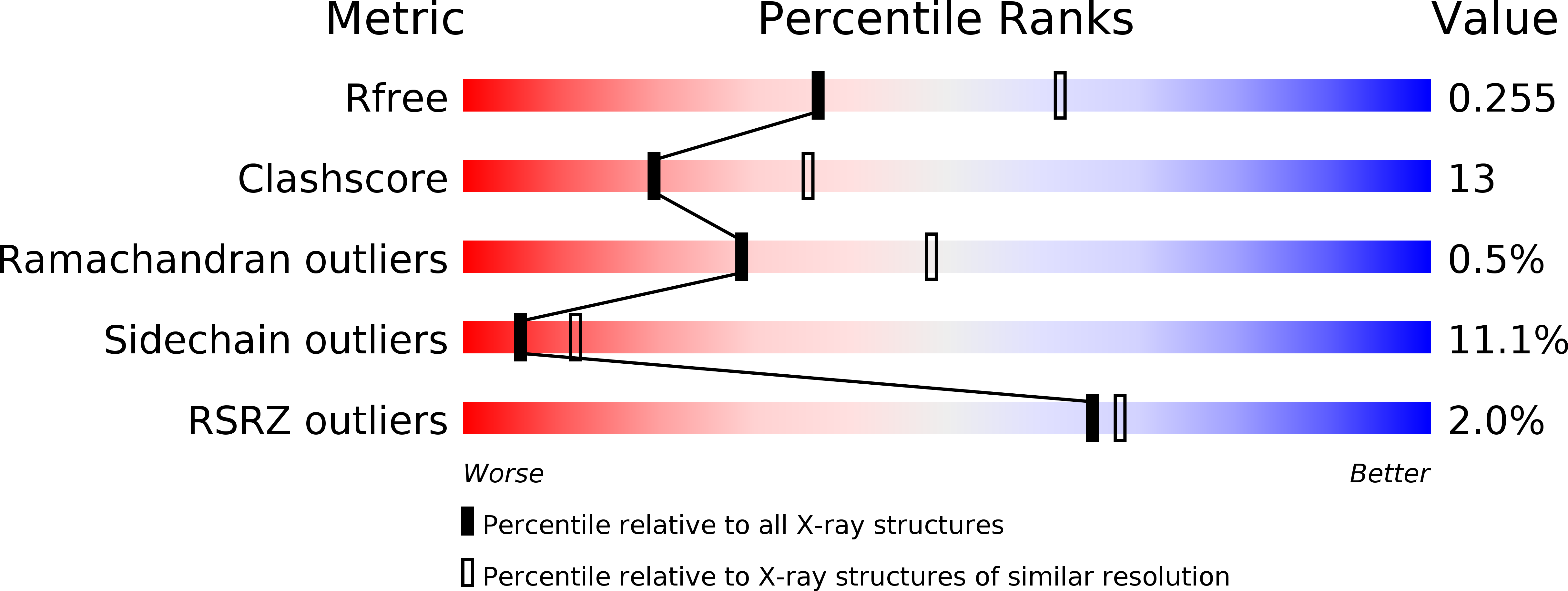
R-Value Free:
0.25
R-Value Work:
0.18
R-Value Observed:
0.18
Space Group:
P 21 21 21