2CVV image
Deposition Date 2005-06-14
Release Date 2006-03-07
Last Version Date 2024-04-03
Entry Detail
PDB ID:
2CVV
Keywords:
Title:
Structures of Yeast Ribonucleotide Reductase I
Biological Source:
Source Organism:
Method Details:
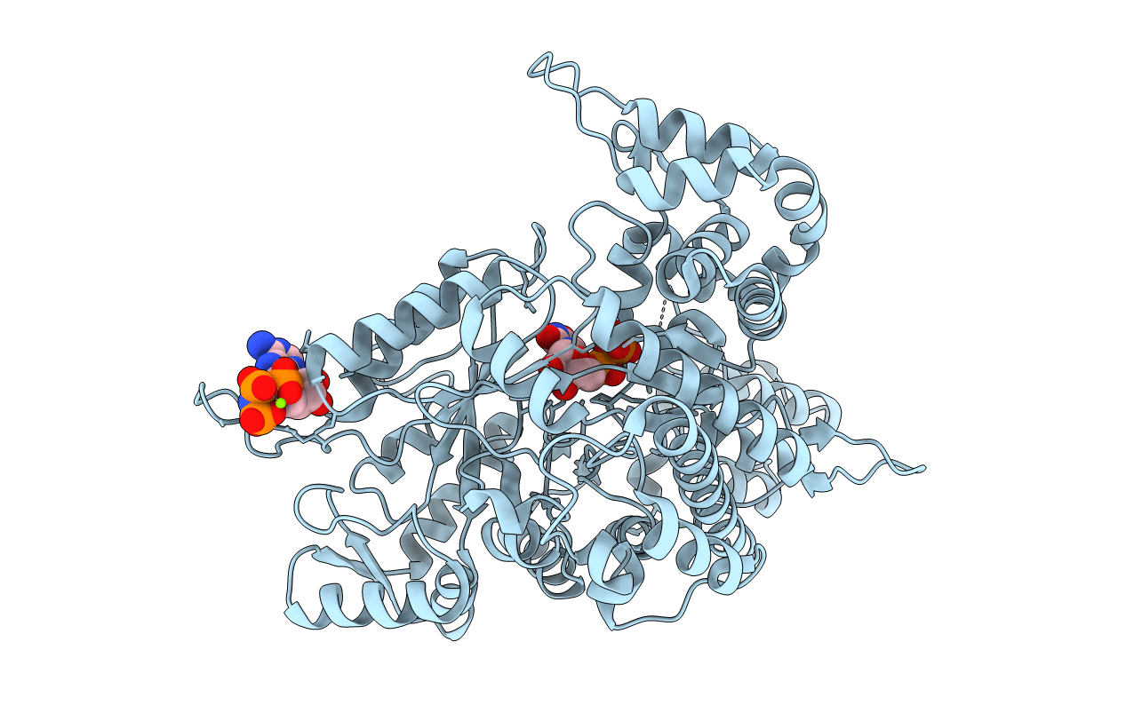
Experimental Method:
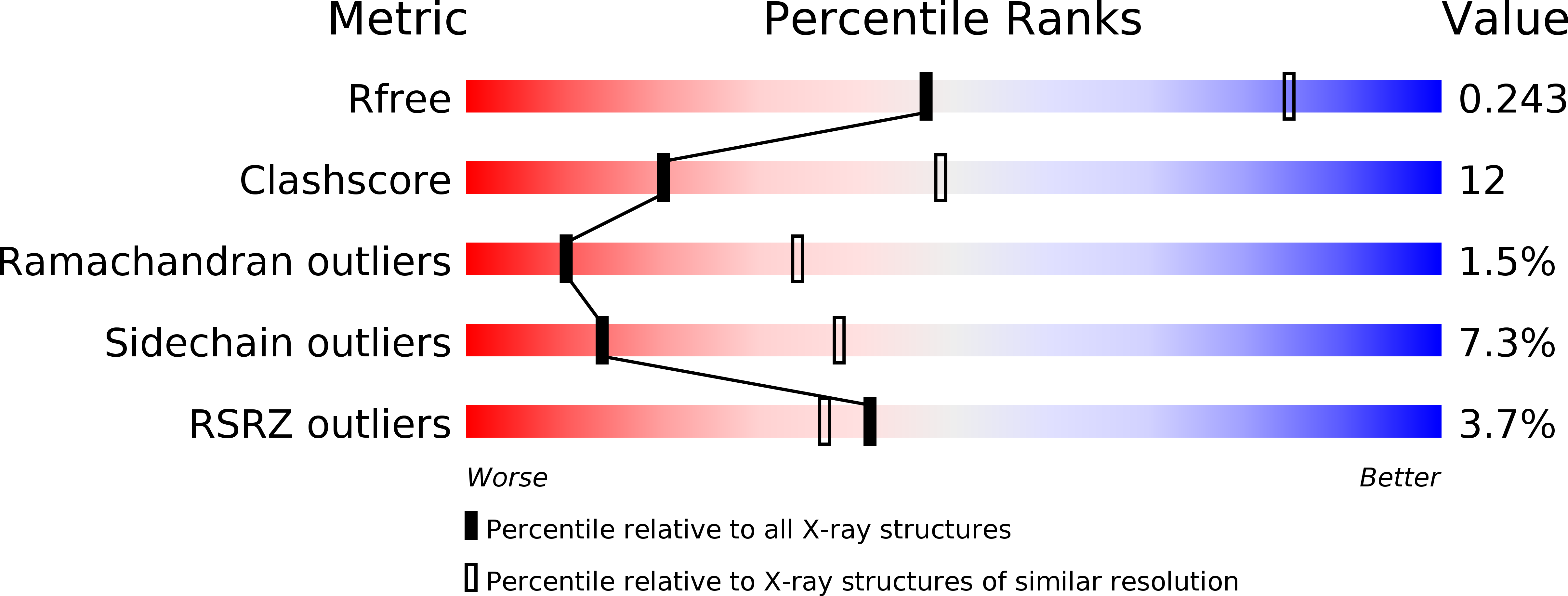
Resolution:
2.90 Å
R-Value Free:
0.24
R-Value Work:
0.17
R-Value Observed:
0.18
Space Group:
P 21 21 2
Macromolecular Entities
Polymer Type:polypeptide(L)
Molecule:Ribonucleoside-diphosphate reductase large chain 1
Gene (Uniprot):RNR1
Chain IDs:A
Chain Length:888
Number of Molecules:1
Biological Source:Saccharomyces cerevisiae
Primary Citation
Structures of eukaryotic ribonucleotide reductase I provide insights into dNTP regulation
Proc.Natl.Acad.Sci.Usa 103 4022 4027 (2006)
PMID: 16537479 DOI: 10.1073/pnas.0600443103

Abstact

Ribonucleotide reductase catalyzes a crucial step in de novo DNA synthesis and is allosterically controlled by relative levels of dNTPs to maintain a balanced pool of deoxynucleoside triphosphates in the cell. In eukaryotes, the enzyme comprises a heterooligomer of alpha(2) and beta(2) subunits. The alpha subunit, Rnr1, contains catalytic and regulatory sites. Here, we report the only x-ray structures of the eukaryotic alpha subunit of ribonucleotide reductase from Saccharomyces cerevisiae. The structures of the apo-, AMPPNP only-, AMPPNP-CDP-, AMPPNP-UDP-, dGTP-ADP- and TTP-GDP-bound complexes give insight into substrate and effector binding and specificity cross-talk. These are Class I structures with the only fully ordered catalytic sites, including loop 2, a stretch of polypeptide that spans specificity and catalytic sites, conferring specificity. Binding of specificity effector rearranges loop 2; in our structures, this rearrangement moves P294, a residue unique to eukaryotes, out of the catalytic site, accommodating substrate binding. Substrate binding further rearranges loop 2. Cross-talk, by which effector binding regulates substrate preference, occurs largely through R293 and Q288 of loop 2, which are analogous to residues in Thermotoga maritima that mediate cross-talk. However loop-2 conformations and residue-substrate interactions differ substantially between yeast and T. maritima. In most effector-substrate complexes, water molecules help mediate substrate-loop 2 interactions. Finally, the substrate ribose binds with its 3' hydroxyl closer than its 2' hydroxyl to C218 of the catalytic redox pair. We also see a conserved water molecule at the catalytic site in all our structures, near the ribose 2' hydroxyl.

Legend

Protein

Chemical

Disease

Primary Citation of related structures