2CUA image
Deposition Date 1999-02-18
Release Date 1999-05-28
Last Version Date 2023-12-27
Entry Detail
PDB ID:
2CUA
Title:
THE CUA DOMAIN OF CYTOCHROME BA3 FROM THERMUS THERMOPHILUS
Biological Source:
Source Organism:
Host Organism:
Method Details:
Experimental Method:
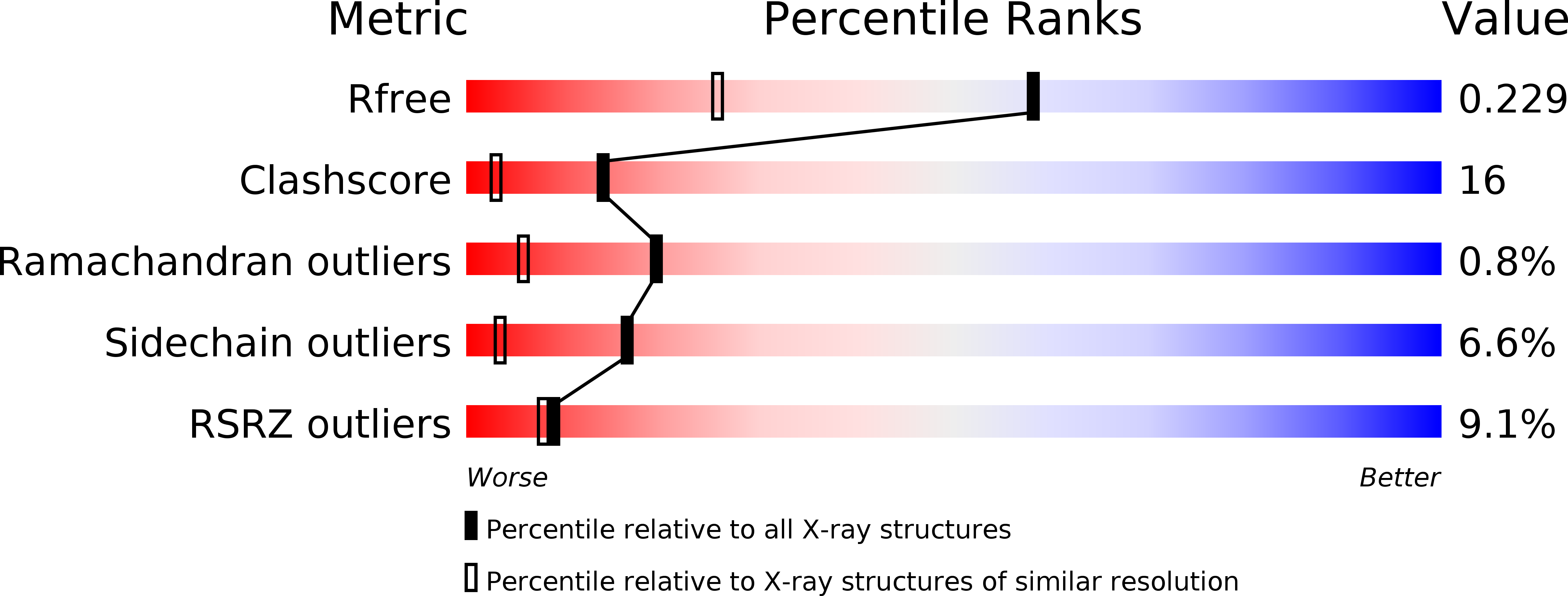
Resolution:
1.60 Å
R-Value Free:
0.29
R-Value Observed:
0.22
Space Group:
P 1 21 1
Macromolecular Entities
Structures with similar UniProt ID
Protein Blast
Polymer Type:polypeptide(L)
Molecule:PROTEIN (CUA)
Gene (Uniprot):cbaB
Chain IDs:A, B
Chain Length:135
Number of Molecules:2
Biological Source:Thermus thermophilus
Primary Citation
The CuA domain of Thermus thermophilus ba3-type cytochrome c oxidase at 1.6 A resolution.
Nat.Struct.Biol. 6 509 516 (1999)
PMID: 10360350 DOI: 10.1038/9274

Abstact

The structure of the CuA-containing, extracellular domain of Thermus thermophilus ba3-type cytochrome c oxidase has been determined to 1.6 A resolution using multiple X-ray wavelength anomalous dispersion (MAD). The Cu2S2 cluster forms a planar rhombus with a copper-copper distance of 2.51 +/- 0.03 A. X-ray absorption fine-structure (EXAFS) studies show that this distance is unchanged by crystallization. The CuA center is asymmetrical; one copper is tetrahedrally coordinated to two bridging cysteine thiolates, one histidine nitrogen and one methionine sulfur, while the other is trigonally coordinated by the two cysteine thiolates and a histidine nitrogen. Combined sequence-structure alignment of amino acid sequences reveals conserved interactions between cytochrome c oxidase subunits I and II.

Legend

Protein

Chemical

Disease

Primary Citation of related structures