2CSB image
Deposition Date 2005-05-21
Release Date 2006-01-31
Last Version Date 2024-11-20
Entry Detail
PDB ID:
2CSB
Keywords:
Title:
Crystal structure of Topoisomerase V from Methanopyrus kandleri (61 kDa fragment)
Biological Source:
Source Organism:
Method Details:
Experimental Method:
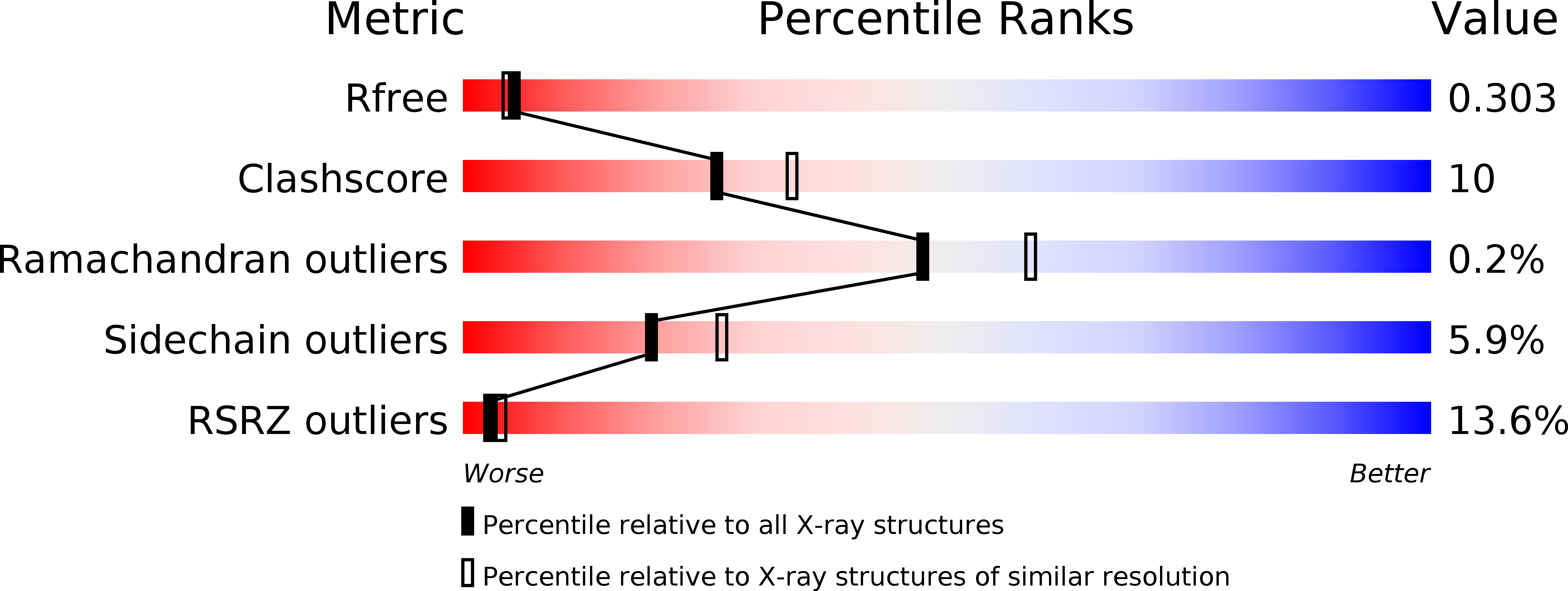
Resolution:
2.30 Å
R-Value Free:
0.29
R-Value Work:
0.21
R-Value Observed:
0.22
Space Group:
P 21 21 21
Macromolecular Entities
Structures with similar UniProt ID
Protein Blast
Polymer Type:polypeptide(L)
Molecule:Topoisomerase V
Chain IDs:A, B
Chain Length:519
Number of Molecules:2
Biological Source:Methanopyrus kandleri
Modified Residue
Compound ID Chain ID Parent Comp ID Details 2D Image
MSE A MET SELENOMETHIONINE
Ligand Molecules
Primary Citation
Structure of the N-terminal fragment of topoisomerase V reveals a new family of topoisomerases
Embo J. 25 398 408 (2006)
PMID: 16395333 DOI: 10.1038/sj.emboj.7600922

Abstact

Topoisomerases are involved in controlling and maintaining the topology of DNA and are present in all kingdoms of life. Unlike all other types of topoisomerases, similar type IB enzymes have only been identified in bacteria and eukarya. The only putative type IB topoisomerase in archaea is represented by Methanopyrus kandleri topoisomerase V. Despite several common functional characteristics, topoisomerase V shows no sequence similarity to other members of the same type. The structure of the 61 kDa N-terminal fragment of topoisomerase V reveals no structural similarity to other topoisomerases. Furthermore, the structure of the active site region is different, suggesting no conservation in the cleavage and religation mechanism. Additionally, the active site is buried, indicating the need of a conformational change for activity. The presence of a topoisomerase in archaea with a unique structure suggests the evolution of a separate mechanism to alter DNA.

Legend

Protein

Chemical

Disease

Primary Citation of related structures