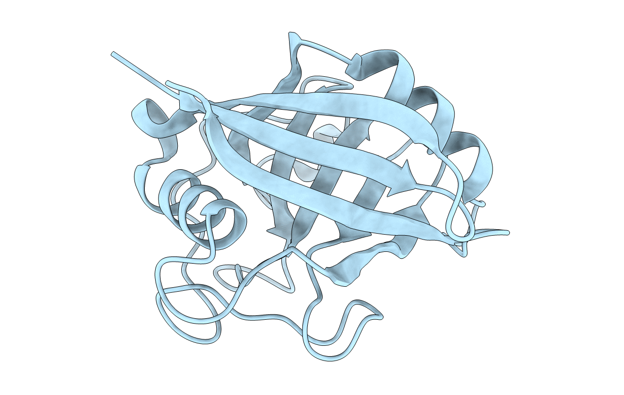
2CPL image
Deposition Date 1992-06-30
Release Date 1993-10-31
Last Version Date 2024-02-14
Entry Detail
PDB ID:
2CPL
Title:
SIMILARITIES AND DIFFERENCES BETWEEN HUMAN CYCLOPHILIN A AND OTHER BETA-BARREL STRUCTURES. STRUCTURAL REFINEMENT AT 1.63 ANGSTROMS RESOLUTION
Biological Source:
Source Organism:
Homo sapiens (Taxon ID: 9606)
Method Details:
Experimental Method:
Resolution:
1.63 Å
R-Value Work:
0.18
R-Value Observed:
0.18
Space Group:
P 21 21 21
Macromolecular Entities
Polymer Type:polypeptide(L)
Molecule:CYCLOPHILIN A
Gene (Uniprot):PPIA
Chain IDs:A
Chain Length:165
Number of Molecules:1
Biological Source:Homo sapiens
Primary Citation
Similarities and differences between human cyclophilin A and other beta-barrel structures. Structural refinement at 1.63 A resolution.
J.Mol.Biol. 228 539 550 (1992)
PMID: 1453463 DOI: 10.1016/0022-2836(92)90841-7

Abstact

The structure of the unligated recombinant human cyclophilin A (CyP A) has been refined to an R-factor of 0.18 at 1.63 A resolution. The root-mean-squared deviations of the refined structure are 0.013 A and 2.50 degrees from ideal geometries of bond length and bond angle, respectively. Eight antiparallel beta-strands of CyP A form a right-handed beta-barrel. The structure of CyP A is compared with other members in the antiparallel eight-stranded beta-barrel family and with the parallel eight-stranded alpha/beta barrels. Although all known eight-stranded barrels are right-handed, the tilted angle of the strands against the barrel axis varies from 45 degrees for retinol binding protein and 49 degrees for CyP A to 70 degrees for superoxide dismutase. As a result, the beta-barrel of CyP A is not completely superimposable with other members of beta-barrels. The structure of CyP A has a unique topology, distinct from other members in the beta-barrel family. In addition, CyP A is a closed beta-barrel so that neither the immunosuppressive drug cyclosporin A (CsA) nor the proline-containing substrate can bind to the hydrophobic core of the CyP A barrel, while the hydrophobic core of most other barrels is open for ligation. These observations probably indicate that CyP A is neither functionally nor evolutionally related to other beta-barrel structures. Details of interactions between solvent molecules and the active site residues of CyP A are illustrated. A water-co-operated mechanism, where the cis<-->trans isomerization might possibly consist of (1) transition of the prolyl bond and (2) release of N or C-terminal residues of substrate from CyP, is addressed. The refined structure reveals no disulfide bridges in CyP A. Cys115 is near the CsA site, but unlikely to be directly involved in CsA binding because of steric hindrance from Thr119 and Leu122. This geometry probably rules out any mechanisms involving a tetrahedral intermediate formed between cysteine and substrate during cis<-->trans isomerization.

Legend

Protein

Chemical

Disease

Primary Citation of related structures