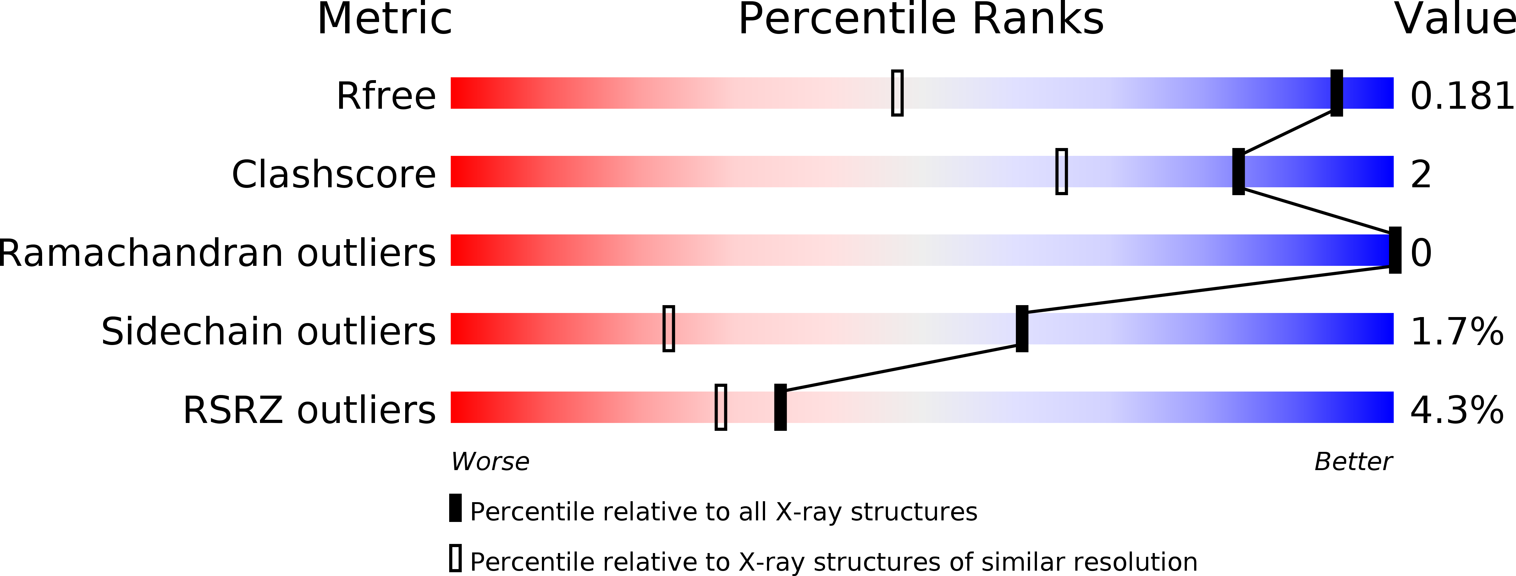
2COV image
Deposition Date 2005-05-18
Release Date 2005-09-13
Last Version Date 2024-11-20
Entry Detail
PDB ID:
2COV
Title:
Crystal structure of CBM31 from beta-1,3-xylanase
Biological Source:
Source Organism:
Host Organism:
Method Details:
Experimental Method:
Resolution:
1.25 Å
R-Value Free:
0.17
R-Value Work:
0.14
R-Value Observed:
0.15
Space Group:
P 21 21 21
Macromolecular Entities
Polymer Type:polypeptide(L)
Molecule:beta-1,3-xylanase
Gene (Uniprot):txyA
Chain IDs:A (auth: D), B (auth: E), C (auth: F), D (auth: G), E (auth: H), F (auth: I)
Chain Length:104
Number of Molecules:6
Biological Source:Alcaligenes sp.
Primary Citation
The first crystal structure of a family 31 carbohydrate-binding module with affinity to beta-1,3-xylan
Febs Lett. 579 4324 4328 (2005)
PMID: 16061225 DOI: 10.1016/j.febslet.2005.06.062

Abstact

Here, we present the crystal structure of the family 31 carbohydrate-binding module (CBM) of beta-1,3-xylanase from Alcaligenes sp. strain XY-234 (AlcCBM31) determined at a resolution of 1.25A. The AlcCBM31 shows affinity with only beta-1,3-xylan. The AlcCBM31 molecule makes a beta-sandwich structure composed of eight beta-strands with a typical immunoglobulin fold and contains two intra-molecular disulfide bonds. The folding topology of AlcCBM31 differs from that of the large majority of other CBMs, in which eight beta-strands comprise a beta-sandwich structure with a typical jelly-roll fold. AlcCBM31 shows structural similarity with CBM structures of family 34 and family 9, which also adopt structures based on immunoglobulin folds.

Legend

Protein

Chemical

Disease

Primary Citation of related structures