2COL image
Deposition Date 2005-05-18
Release Date 2006-01-24
Last Version Date 2024-03-13
Entry Detail
PDB ID:
2COL
Title:
Crystal structure analysis of CyaA/C-Cam with Pyrophosphate
Biological Source:
Source Organism:
Host Organism:
Method Details:
Experimental Method:
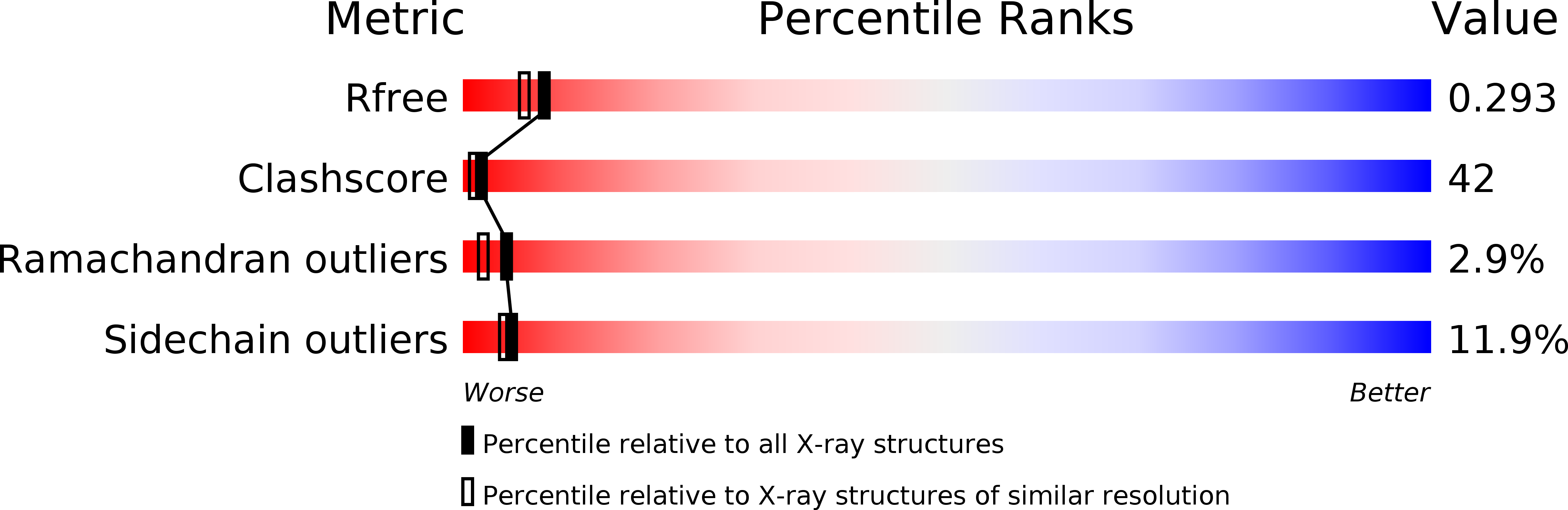
Resolution:
2.20 Å
R-Value Free:
0.29
R-Value Work:
0.24
R-Value Observed:
0.24
Space Group:
P 41 21 2
Macromolecular Entities
Structures with similar UniProt ID
Protein Blast
Polymer Type:polypeptide(L)
Molecule:Bifunctional hemolysin-adenylate cyclase
Chain IDs:A
Chain Length:356
Number of Molecules:1
Biological Source:Bordetella pertussis
Structures with similar UniProt ID
Protein Blast
Polymer Type:polypeptide(L)
Molecule:Calmodulin
Chain IDs:B
Chain Length:67
Number of Molecules:1
Biological Source:Xenopus laevis
Primary Citation
Structural basis for the interaction of Bordetella pertussis adenylyl cyclase toxin with calmodulin
Embo J. 24 3190 3201 (2005)
PMID: 16138079 DOI: 10.1038/sj.emboj.7600800

Abstact

CyaA is crucial for colonization by Bordetella pertussis, the etiologic agent of whooping cough. Here we report crystal structures of the adenylyl cyclase domain (ACD) of CyaA with the C-terminal domain of calmodulin. Four discrete regions of CyaA bind calcium-loaded calmodulin with a large buried contact surface. Of those, a tryptophan residue (W242) at an alpha-helix of CyaA makes extensive contacts with the calcium-induced, hydrophobic pocket of calmodulin. Mutagenic analyses show that all four regions of CyaA contribute to calmodulin binding and the calmodulin-induced conformational change of CyaA is crucial for catalytic activation. A crystal structure of CyaA-calmodulin with adefovir diphosphate, the metabolite of an approved antiviral drug, reveals the location of catalytic site of CyaA and how adefovir diphosphate tightly binds CyaA. The ACD of CyaA shares a similar structure and mechanism of activation with anthrax edema factor (EF). However, the interactions of CyaA with calmodulin completely diverge from those of EF. This provides molecular details of how two structurally homologous bacterial toxins evolved divergently to bind calmodulin, an evolutionarily conserved calcium sensor.

Legend

Protein

Chemical

Disease

Primary Citation of related structures