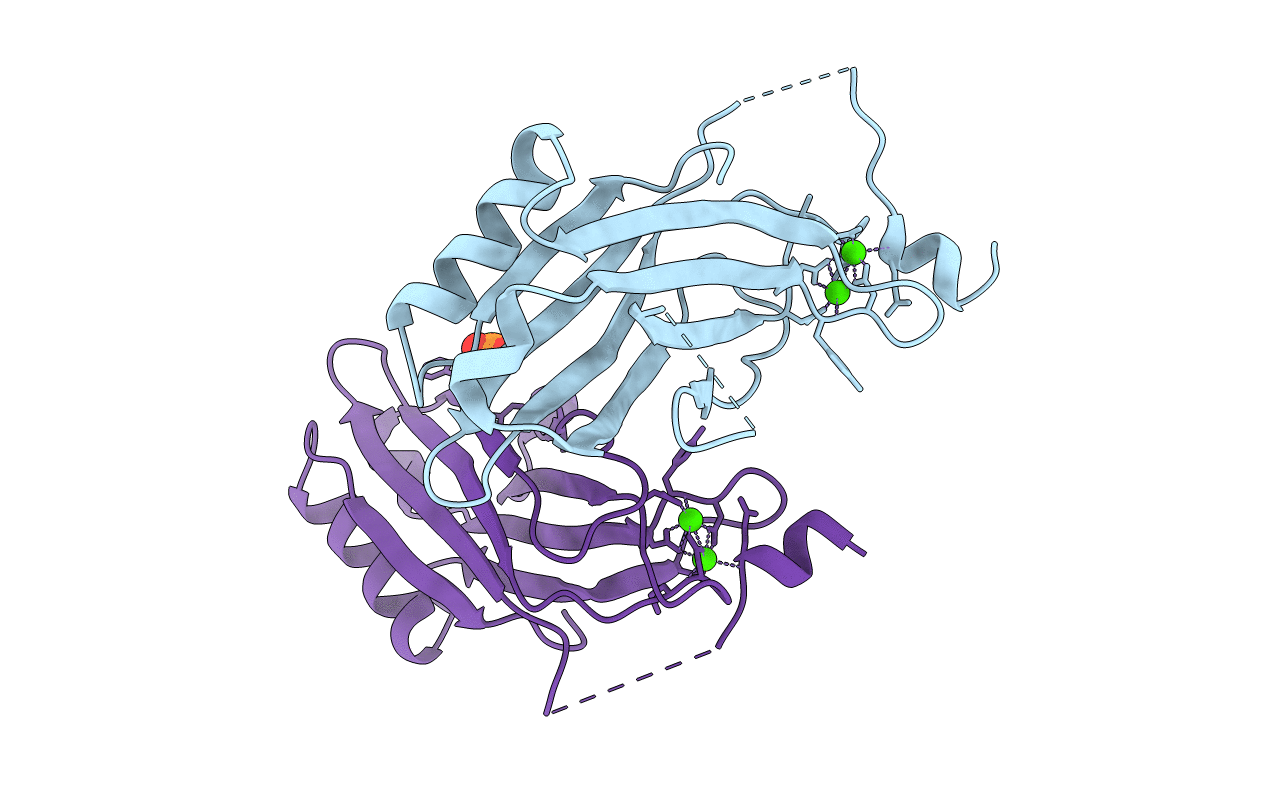
2CM6 image
Entry Detail
PDB ID:
2CM6
Title:
crystal structure of the C2B domain of rabphilin3A
Biological Source:
Source Organism:
Host Organism:
PDB Version:
Deposition Date:
2006-05-04
Release Date:
2006-12-04
Method Details:
Experimental Method:
Resolution:
1.85 Å
R-Value Free:
0.26
R-Value Observed:
0.19
Space Group:
P 1 21 1
Macromolecular Entities
Polymer Type:polypeptide(L)
Description:RABPHILIN-3A
Chain IDs:A, B
Chain Length:166
Number of Molecules:2
Biological Source:RATTUS NORVEGICUS
Primary Citation
The C2A-C2B Linker Defines the High Affinity Ca2+ Binding Mode of Rabphilin-3A.
J.Biol.Chem. 282 5015 ? (2007)
PMID: 17166855 DOI: 10.1074/JBC.M606746200

Abstact

The Ca(2+) binding properties of C2 domains are essential for the function of their host proteins. We present here the first crystal structures showing an unexpected Ca(2+) binding mode of the C2B domain of rabphilin-3A in atomic detail. Acidic residues from the linker region between the C2A and C2B domains of rabphilin-3A interact with the Ca(2+)-binding region of the C2B domain. Because of these interactions, the coordination sphere of the two bound Ca(2+) ions is almost complete. Mutation of these acidic residues to alanine resulted in a 10-fold decrease in the intrinsic Ca(2+) binding affinity of the C2B domain. Using NMR spectroscopy, we show that this interaction occurred only in the Ca(2+)-bound state of the C2B domain. In addition, this Ca(2+) binding mode was maintained in the C2 domain tandem fragment. In NMR-based liposome binding assays, the linker was not released upon phospholipid binding. Therefore, this unprecedented Ca(2+) binding mode not only shows how a C2 domain increases its intrinsic Ca(2+) affinity, but also provides the structural base for an atypical protein-Ca(2+)-phospholipid binding mode of rabphilin-3A.

Legend

Protein

Chemical

Disease

Primary Citation of related structures