Deposition Date
2006-04-25
Release Date
2006-10-25
Last Version Date
2024-10-16
Entry Detail
PDB ID:
2CL2
Keywords:
Title:
Endo-1,3(4)-beta-glucanase from Phanerochaete chrysosporium, solved using native sulfur SAD, exhibiting intact heptasaccharide glycosylation
Biological Source:
Source Organism(s):
PHANEROCHAETE CHRYSOSPORIUM (Taxon ID: 5306)
Expression System(s):
Method Details:
Experimental Method:
Resolution:
1.35 Å
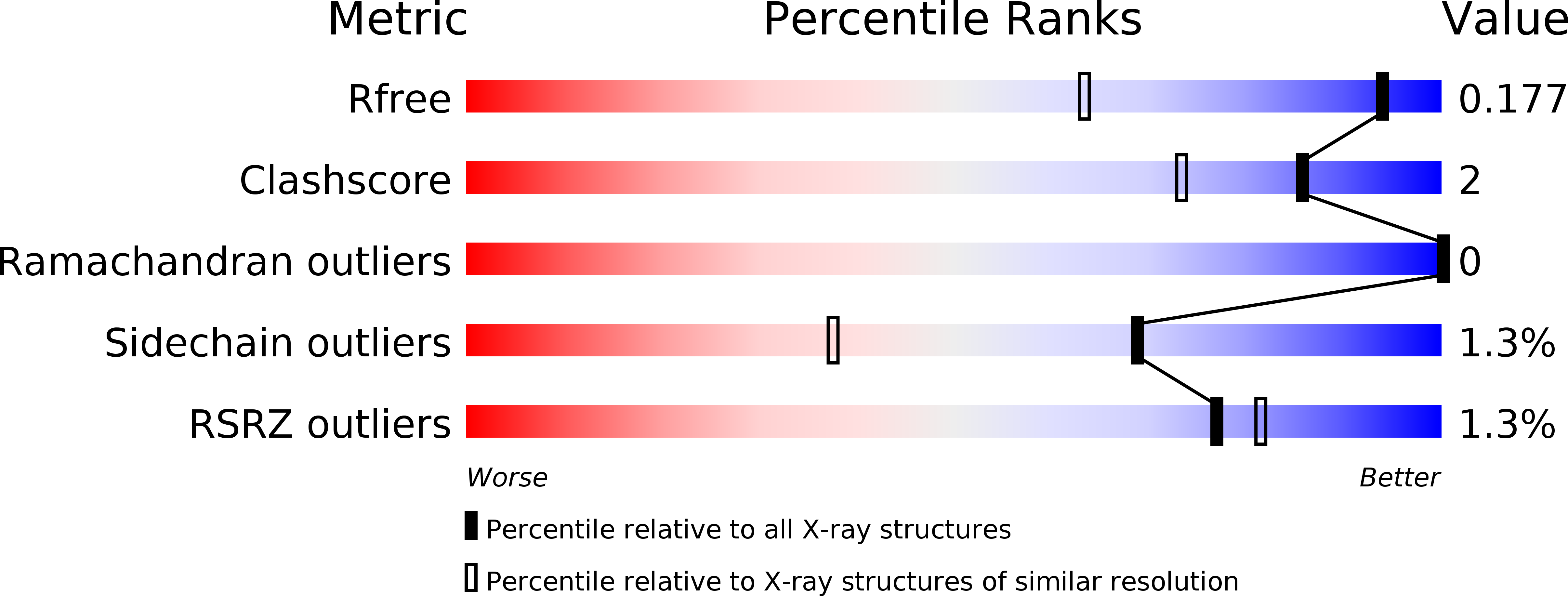
R-Value Free:
0.16
R-Value Work:
0.14
R-Value Observed:
0.14
Space Group:
P 21 21 21