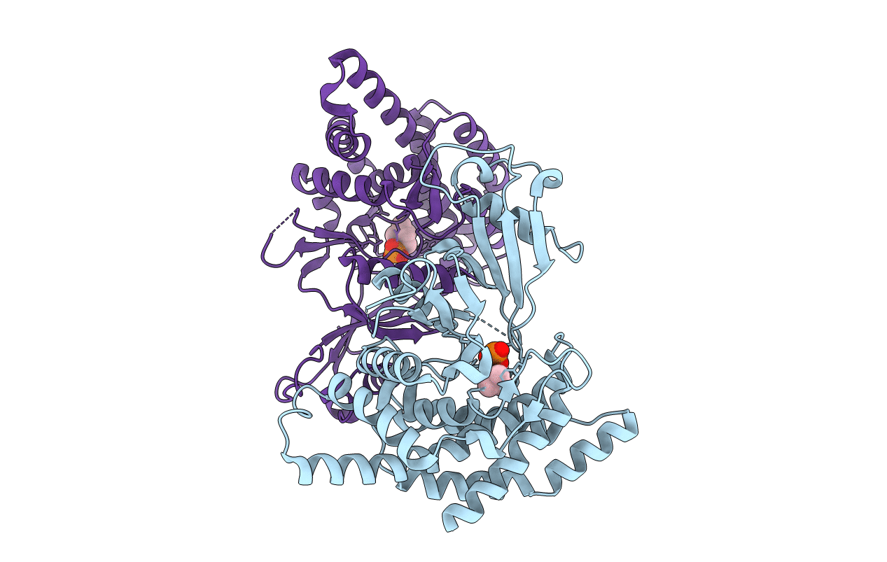
2CKQ image
Entry Detail
PDB ID:
2CKQ
Keywords:
Title:
Crystal structure of Human Choline Kinase alpha 2 in complex with Phosphocholine
Biological Source:
Source Organism:
Host Organism:
PDB Version:
Deposition Date:
2006-04-20
Release Date:
2006-10-04
Method Details:
Experimental Method:
Resolution:
2.40 Å
R-Value Free:
0.25
R-Value Work:
0.21
R-Value Observed:
0.21
Space Group:
P 21 21 21
Macromolecular Entities
Polymer Type:polypeptide(L)
Description:CHOLINE KINASE ALPHA
Chain IDs:A, B
Chain Length:390
Number of Molecules:2
Biological Source:HOMO SAPIENS
Ligand Molecules
Primary Citation
Elucidation of Human Choline Kinase Crystal Structures in Complex with the Products Adp or Phosphocholine.
J.Mol.Biol. 364 136 ? (2006)
PMID: 17007874 DOI: 10.1016/J.JMB.2006.08.084

Abstact

Choline kinase, responsible for the phosphorylation of choline to phosphocholine as the first step of the CDP-choline pathway for the biosynthesis of phosphatidylcholine, has been recognized as a new target for anticancer therapy. Crystal structures of human choline kinase in its apo, ADP and phosphocholine-bound complexes, respectively, reveal the molecular details of the substrate binding sites. ATP binds in a cavity where residues from both the N and C-terminal lobes contribute to form a cleft, while the choline-binding site constitutes a deep hydrophobic groove in the C-terminal domain with a rim composed of negatively charged residues. Upon binding of choline, the enzyme undergoes conformational changes independently affecting the N-terminal domain and the ATP-binding loop. From this structural analysis and comparison with other kinases, and from mutagenesis data on the homologous Caenorhabditis elegans choline kinase, a model of the ternary ADP.phosphocholine complex was built that reveals the molecular basis for the phosphoryl transfer activity of this enzyme.

Legend

Protein

Chemical

Disease

Primary Citation of related structures