Deposition Date
2006-04-09
Release Date
2006-06-27
Last Version Date
2024-11-13
Entry Detail
PDB ID:
2CJX
Keywords:
Title:
Extended substrate recognition in caspase-3 revealed by high resolution X-ray structure analysis
Biological Source:
Source Organism(s):
HOMO SAPIENS (Taxon ID: 9606)
SYNTHETIC CONSTRUCT (Taxon ID: 32630)
SYNTHETIC CONSTRUCT (Taxon ID: 32630)
Expression System(s):
Method Details:
Experimental Method:
Resolution:
1.70 Å
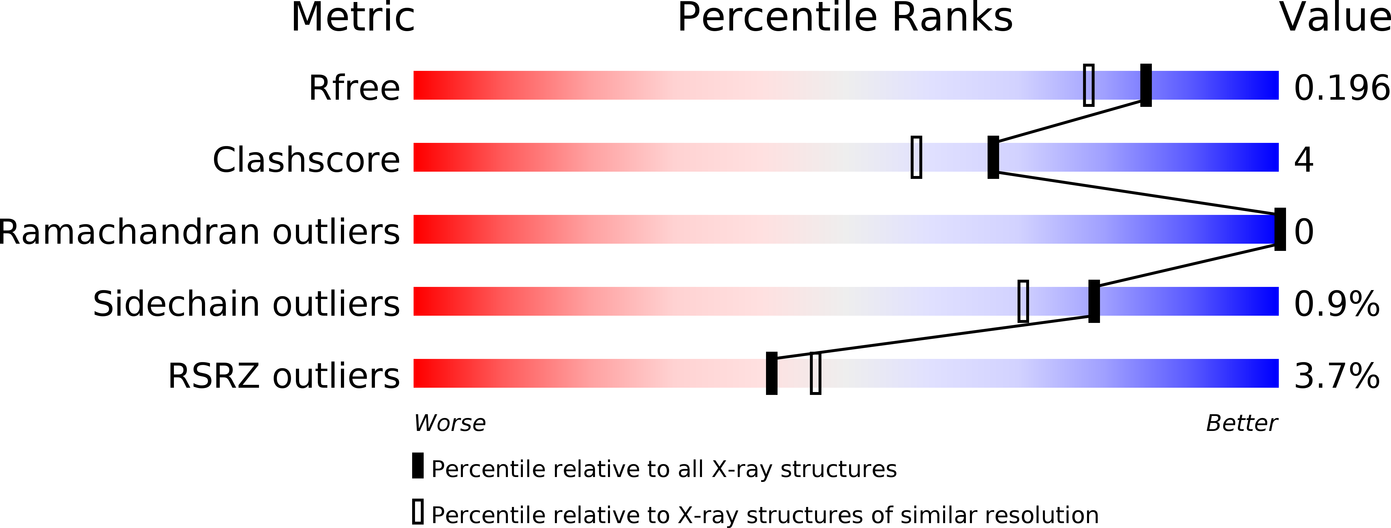
R-Value Free:
0.20
R-Value Work:
0.18
R-Value Observed:
0.18
Space Group:
I 2 2 2