2CHT image
Entry Detail
PDB ID:
2CHT
Keywords:
Title:
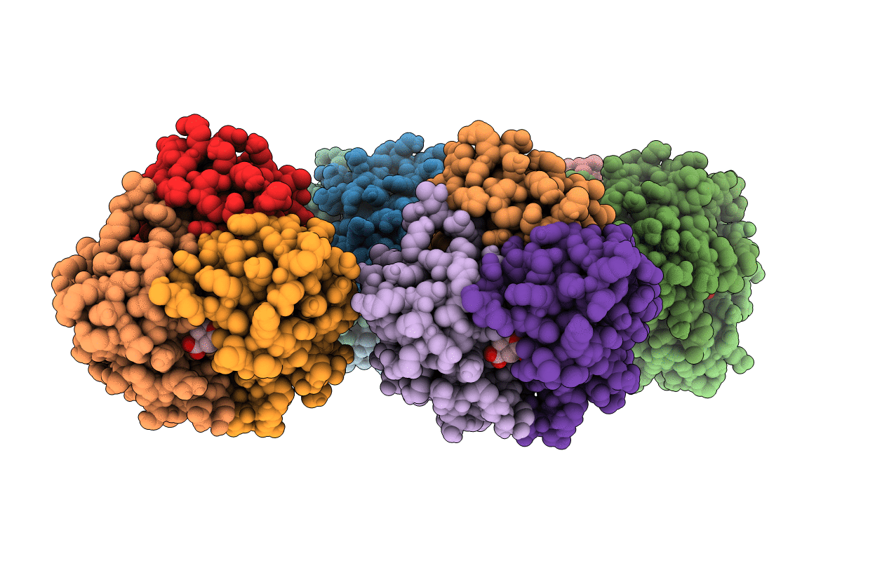
CRYSTAL STRUCTURES OF THE MONOFUNCTIONAL CHORISMATE MUTASE FROM BACILLUS SUBTILIS AND ITS COMPLEX WITH A TRANSITION STATE ANALOG
Biological Source:
Source Organism:
PDB Version:
Deposition Date:
1994-04-08
Release Date:
1994-07-31
Method Details:
Experimental Method:
Resolution:
2.20 Å
R-Value Work:
0.18
R-Value Observed:
0.18
Space Group:
P 1 21 1
Macromolecular Entities
Polymer Type:polypeptide(L)
Description:CHORISMATE MUTASE
Chain IDs:A, B, C, D, E, F, G, H, I, J, K, L
Chain Length:127
Number of Molecules:12
Biological Source:Bacillus subtilis
Ligand Molecules
Primary Citation
Crystal structures of the monofunctional chorismate mutase from Bacillus subtilis and its complex with a transition state analog.
Proc.Natl.Acad.Sci.USA 90 8600 8603 (1993)
PMID: 8378335 DOI: 10.1073/pnas.90.18.8600

Abstact

We have solved the structure of a chorismate mutase (chorismate pyruvatemutase, EC 5.4.99.5), the 1.9-A crystal structure of the monofunctional enzyme from Bacillus subtilis. The structure determination process was an unusual one, involving 12 monomers of the enzyme in the asymmetric unit. This structure was solved by the multiple isomorphous replacement method with partial structure phase combination and molecular averaging. The final model, which includes 1380 residues and 522 water molecules in an asymmetric unit, has been refined at 1.9 A and the current crystallographic R value is 0.201. The B. subtilis chorismate mutase is a homotrimer, with beta-sheets from each monomer packing to form the core of a pseudo-alpha beta-barrel with helices on the outside of the trimer. In addition, the active sites have been located by using data from a complex with an endo-oxabicyclic inhibitor that mimics the transition state of the reaction. The structure of this complex has been refined to 2.2 A with a current R value of 0.182 for a model that includes 1388 residues, 12 inhibitor molecules, and 530 water molecules in the asymmetric unit. In each trimer, three equivalent active sites are located at the interfaces of two adjacent subunits.

Legend

Protein

Chemical

Disease

Primary Citation of related structures