2CGH image
Deposition Date 2006-03-06
Release Date 2007-05-01
Last Version Date 2023-12-13
Entry Detail
PDB ID:
2CGH
Keywords:
Title:
crystal structure of biotin ligase from Mycobacterium tuberculosis
Biological Source:
Source Organism:
Method Details:
Experimental Method:
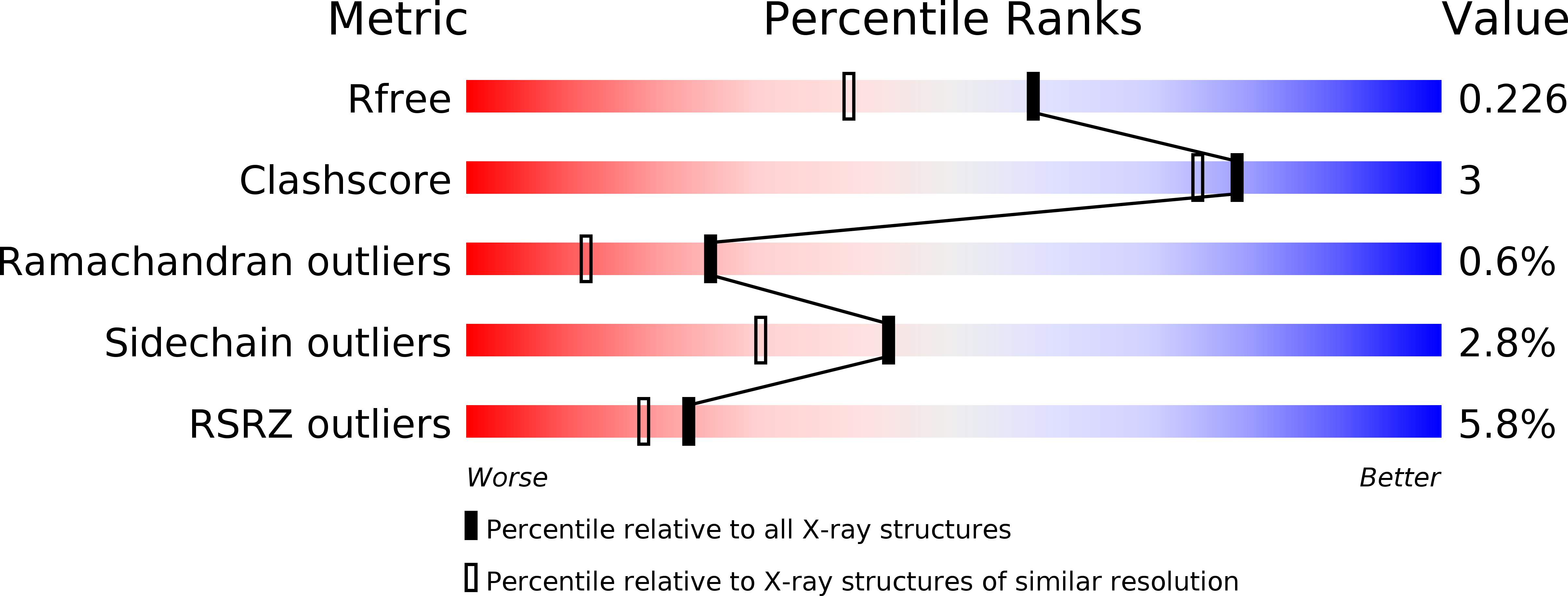
Resolution:
1.80 Å
R-Value Free:
0.22
R-Value Work:
0.18
R-Value Observed:
0.18
Space Group:
P 21 21 21
Macromolecular Entities
Polymer Type:polypeptide(L)
Molecule:BIOTIN LIGASE
Gene (Uniprot):birA
Chain IDs:A, B
Chain Length:268
Number of Molecules:2
Biological Source:MYCOBACTERIUM TUBERCULOSIS
Primary Citation
Active Site Conformational Changes Upon Reaction Intermediate Biotinyl-5'-AMP Binding in Biotin Protein Ligase from Mycobacterium Tuberculosis.
Protein Sci. 23 932 ? (2014)
PMID: 24723382 DOI: 10.1002/PRO.2475

Abstact

Protein biotinylation, a rare form of post-translational modification, is found in enzymes required for lipid biosynthesis. In mycobacteria, this process is essential for the formation of their complex and distinct cell wall and has become a focal point of drug discovery approaches. The enzyme responsible for this process, biotin protein ligase, substantially varies in different species in terms of overall structural organization, regulation of function and substrate specificity. To advance the understanding of the molecular mechanism of biotinylation in Mycobacterium tuberculosis we have biochemically and structurally characterized the corresponding enzyme. We report the high-resolution crystal structures of the apo-form and reaction intermediate biotinyl-5'-AMP-bound form of M. tuberculosis biotin protein ligase. Binding of the reaction intermediate leads to clear disorder-to-order transitions. We show that a conserved lysine, Lys138, in the active site is essential for biotinylation.

Legend

Protein

Chemical

Disease

Primary Citation of related structures