Deposition Date
2006-02-27
Release Date
2007-02-27
Last Version Date
2024-11-13
Entry Detail
PDB ID:
2CG6
Keywords:
Title:
Second and third fibronectin type I module pair (crystal form I).
Biological Source:
Source Organism(s):
HOMO SAPIENS (Taxon ID: 9606)
Expression System(s):
Method Details:
Experimental Method:
Resolution:
1.55 Å
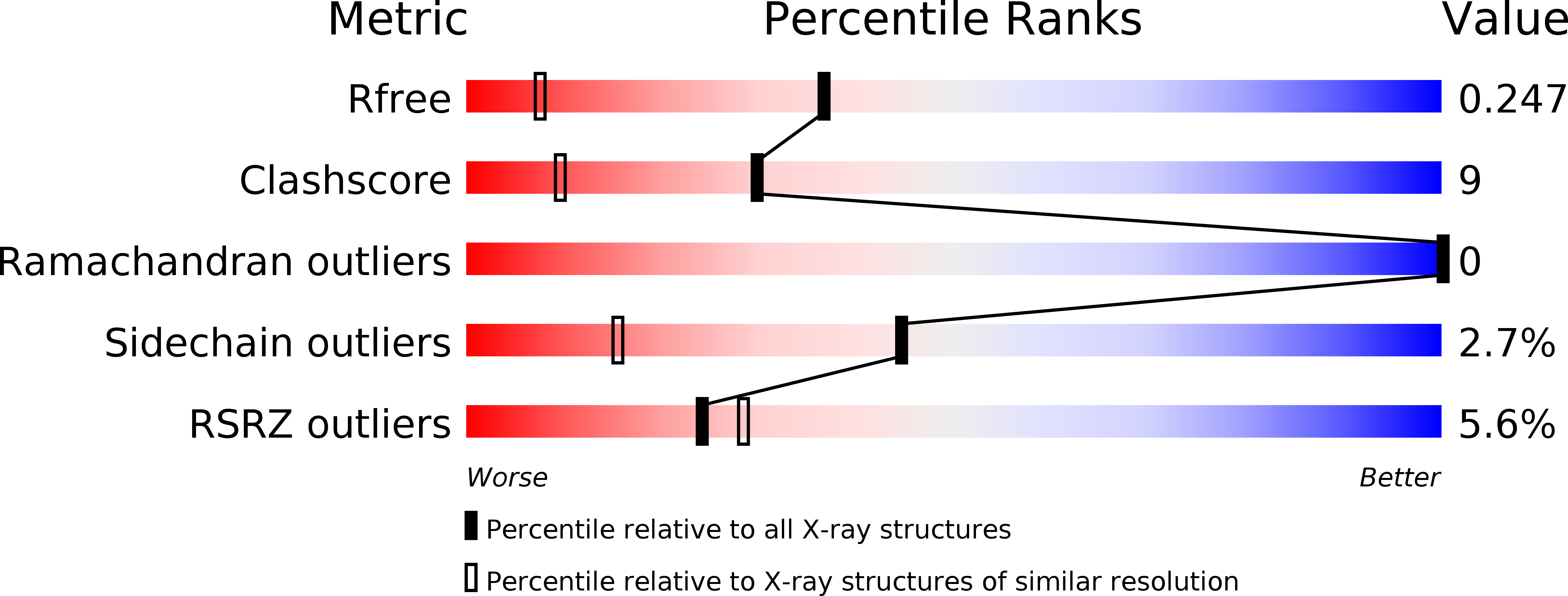
R-Value Free:
0.23
R-Value Work:
0.18
R-Value Observed:
0.19
Space Group:
P 41 21 2