Deposition Date
2006-02-20
Release Date
2006-02-22
Last Version Date
2023-12-13
Entry Detail
PDB ID:
2CFE
Keywords:
Title:
The 1.5 A crystal structure of the Malassezia sympodialis Mala s 6 allergen, a member of the cyclophilin pan-allergen family
Biological Source:
Source Organism(s):
MALASSEZIA SYMPODIALIS (Taxon ID: 76777)
Expression System(s):
Method Details:
Experimental Method:
Resolution:
1.50 Å
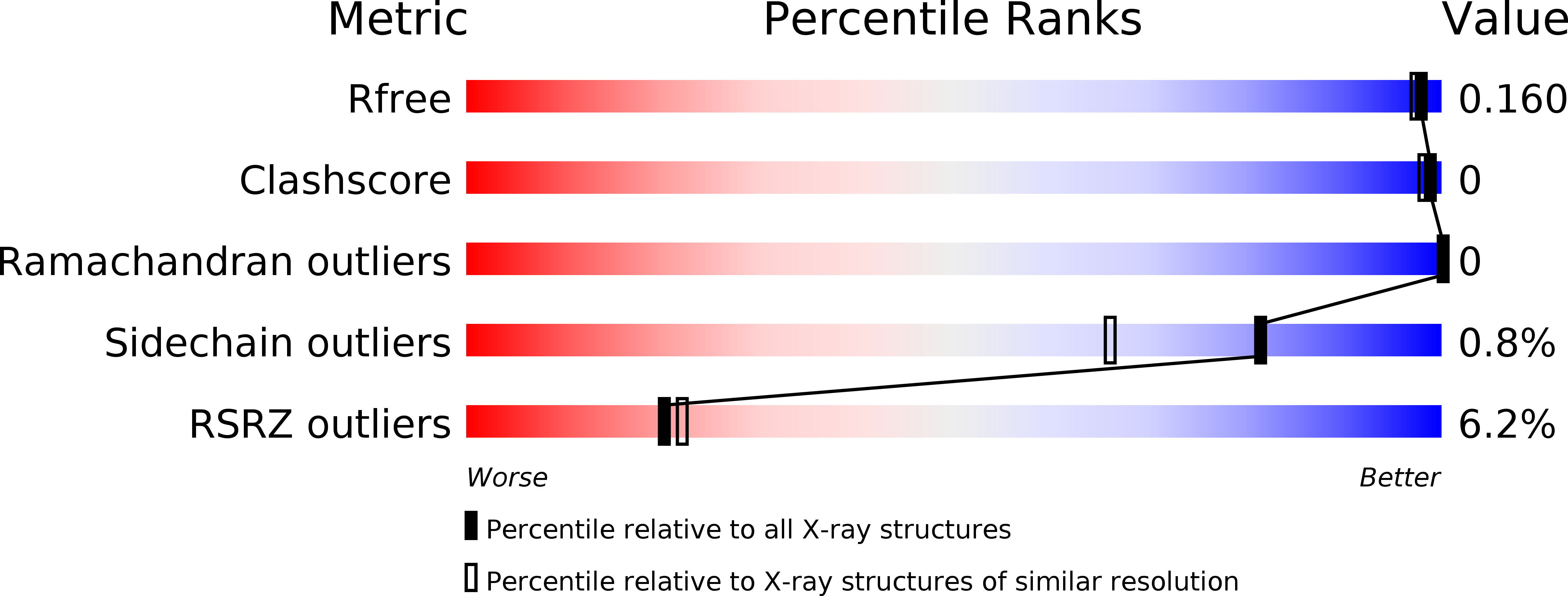
R-Value Free:
0.14
R-Value Work:
0.14
R-Value Observed:
0.14
Space Group:
P 41 21 2