2CC3 image
Deposition Date 2006-01-11
Release Date 2006-01-30
Last Version Date 2023-12-13
Entry Detail
PDB ID:
2CC3
Title:
Structure of Agrobacterium tumefaciens VirB8 protein
Biological Source:
Source Organism:
Host Organism:
Method Details:
Experimental Method:
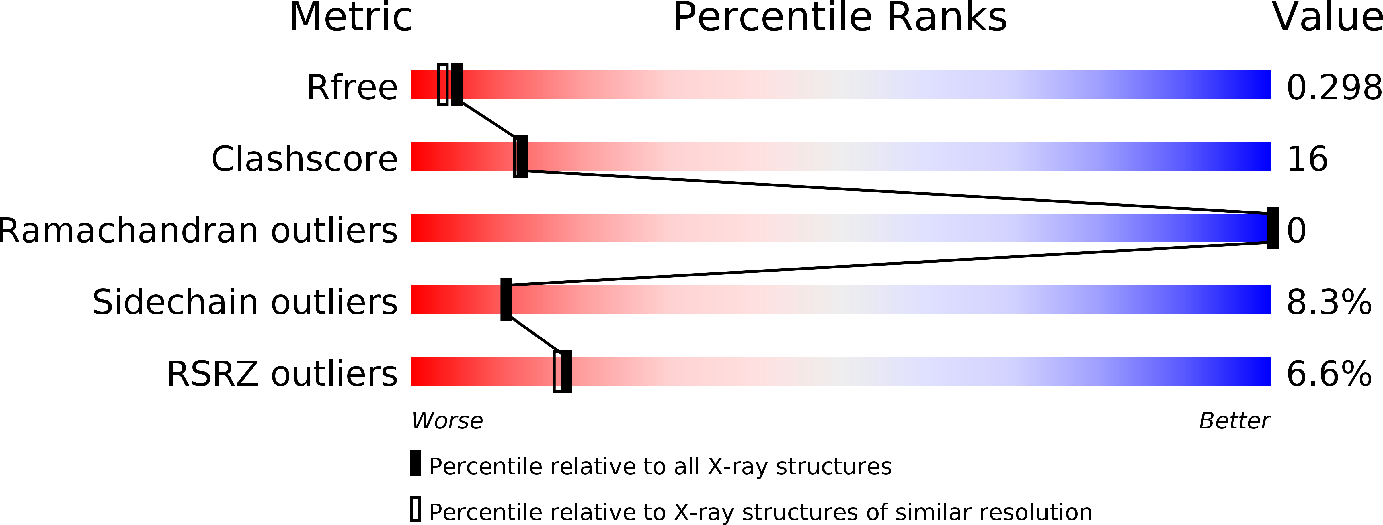
Resolution:
2.20 Å
R-Value Free:
0.30
R-Value Work:
0.23
R-Value Observed:
0.23
Space Group:
C 1 2 1
Macromolecular Entities
Polymer Type:polypeptide(L)
Molecule:PROTEIN VIRB8
Gene (Uniprot):virB8
Chain IDs:A, B
Chain Length:150
Number of Molecules:2
Biological Source:AGROBACTERIUM TUMEFACIENS
Ligand Molecules
Primary Citation
Agrobacterium Tumefaciens Virb8 Structure Reveals Potential Protein-Protein Interactions Sites.
Proc.Natl.Acad.Sci.USA 103 2582 ? (2006)
PMID: 16481621 DOI: 10.1073/PNAS.0511216103

Abstact

Bacterial type IV secretion systems (T4SS) translocate DNA and/or proteins to recipient cells, thus providing a mechanism for conjugative transfer of genetic material and bacterial pathogenesis. Here we describe the first structure of a core component from the archetypal Agrobacterium tumefaciens T4SS: the 2.2-A resolution crystal structure of the VirB8 periplasmic domain (pVirB8(AT)). VirB8 forms a dimer in the crystal, and we identify residues likely important for stabilization of the dimer interface. Structural comparison of pVirB8(AT) with Brucella suis VirB8 confirms that the monomers have a similar fold. In addition, the pVirB8(AT) dimer superimposes very closely on the B. suis VirB8 dimer, supporting the proposal that dimer formation in the crystal reflects self-interactions that are biologically significant. The evolutionary conservation level for each residue was obtained from a data set of 84 VirB8 homologs and projected onto the protein structure to indicate conserved surface patches that likely contact other T4SS proteins.

Legend

Protein

Chemical

Disease

Primary Citation of related structures