2CBT image
Entry Detail
PDB ID:
2CBT
Keywords:
Title:
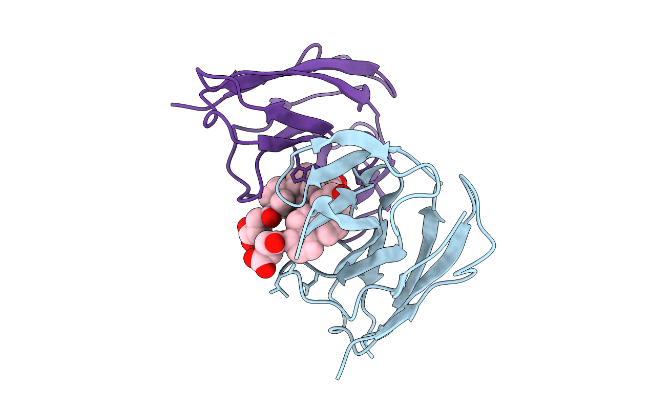
Crystal structure of the neocarzinostatin 4Tes1 mutant bound testosterone hemisuccinate.
Biological Source:
Host Organism:
PDB Version:
Deposition Date:
2006-01-06
Release Date:
2006-03-22
Method Details:
Experimental Method:
Resolution:
2.20 Å
R-Value Free:
0.34
R-Value Observed:
0.21
Space Group:
P 65
Macromolecular Entities
Polymer Type:polypeptide(L)
Description:NEOCARZINOSTATIN
Mutations:YES
Chain IDs:A, B
Chain Length:112
Number of Molecules:2
Biological Source:STREPTOMYCES CARZINOSTATICUS
Ligand Molecules
Primary Citation
Structures of in Vitro Evolved Binding Sites on Neocarzinostatin Scaffold Reveal Unanticipated Evolutionary Pathways.
J.Mol.Biol. 358 455 ? (2006)
PMID: 16529771 DOI: 10.1016/J.JMB.2006.02.002

Abstact

We have recently applied in vitro evolution methods to create in Neocarzinostatin a new binding site for a target molecule unrelated to its natural ligand. The main objective of this work was to solve the structure of some of the selected binders in complex with the target molecule: testosterone. Three proteins (1a.15, 3.24 and 4.1) were chosen as representative members of sequence families that came out of the selection process within different randomization schemes. In order to evaluate ligand-induced conformational adaptation, we also determined the structure of one of the proteins (3.24) in the free and complexed forms. Surprisingly, all these mutants bind not one but two molecules of testosterone in two very different ways. The 3.24 structure revealed that the protein spontaneously evolved in the system to bind two ligand molecules in one single binding crevice. These two binding sites are formed by substituted as well as by non-variable side-chains. The comparison with the free structure shows that only limited structural changes are observed upon ligand binding. The X-ray structures of the complex formed by 1a.15 and 4.1 Neocarzinostatin mutants revealed that the two variants form very similar dimers. These dimers were observed neither for the uncomplexed variants nor for wild-type Neocarzinostatin but were shown here to be induced by ligand binding. Comparison of the three complexed forms clearly suggests that these unanticipated structural responses resulted from the molecular arrangement used for the selection experiments.

Legend

Protein

Chemical

Disease

Primary Citation of related structures