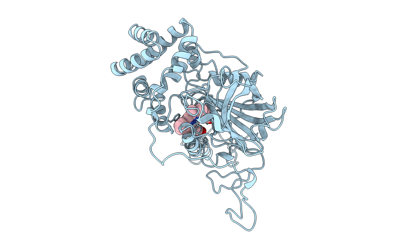
2CAG image
Deposition Date 1996-06-12
Release Date 1996-12-07
Last Version Date 2024-10-23
Entry Detail
PDB ID:
2CAG
Title:
CATALASE COMPOUND II
Biological Source:
Source Organism:
Method Details:
Experimental Method:
Resolution:
2.70 Å
R-Value Free:
0.24
R-Value Work:
0.17
R-Value Observed:
0.17
Space Group:
P 62 2 2
Macromolecular Entities
Structures with similar UniProt ID
Protein Blast
Polymer Type:polypeptide(L)
Molecule:CATALASE COMPOUND II
Gene (Uniprot):katA
Chain IDs:A
Chain Length:484
Number of Molecules:1
Biological Source:Proteus mirabilis
Modified Residue
Compound ID Chain ID Parent Comp ID Details 2D Image
OMT A MET S-DIOXYMETHIONINE
Ligand Molecules
Primary Citation
Ferryl intermediates of catalase captured by time-resolved Weissenberg crystallography and UV-VIS spectroscopy.
Nat.Struct.Biol. 3 951 956 (1996)
PMID: 8901874 DOI: 10.1038/nsb1196-951

Abstact

Various enzymes use semi-stable ferryl intermediates and free radicals during their catalytic cycle, amongst them haem catalases. Structures for two transient intermediates (compounds I and II) of the NADPH-dependent catalase from Proteus mirabilis (PMC) have been determined by time-resolved X-ray crystallography and single crystal microspectrophotometry. The results show the formation and transformation of the ferryl group in the haem, and the unexpected binding of an anion during this reaction at a site distant from the haem.

Legend

Protein

Chemical

Disease

Primary Citation of related structures