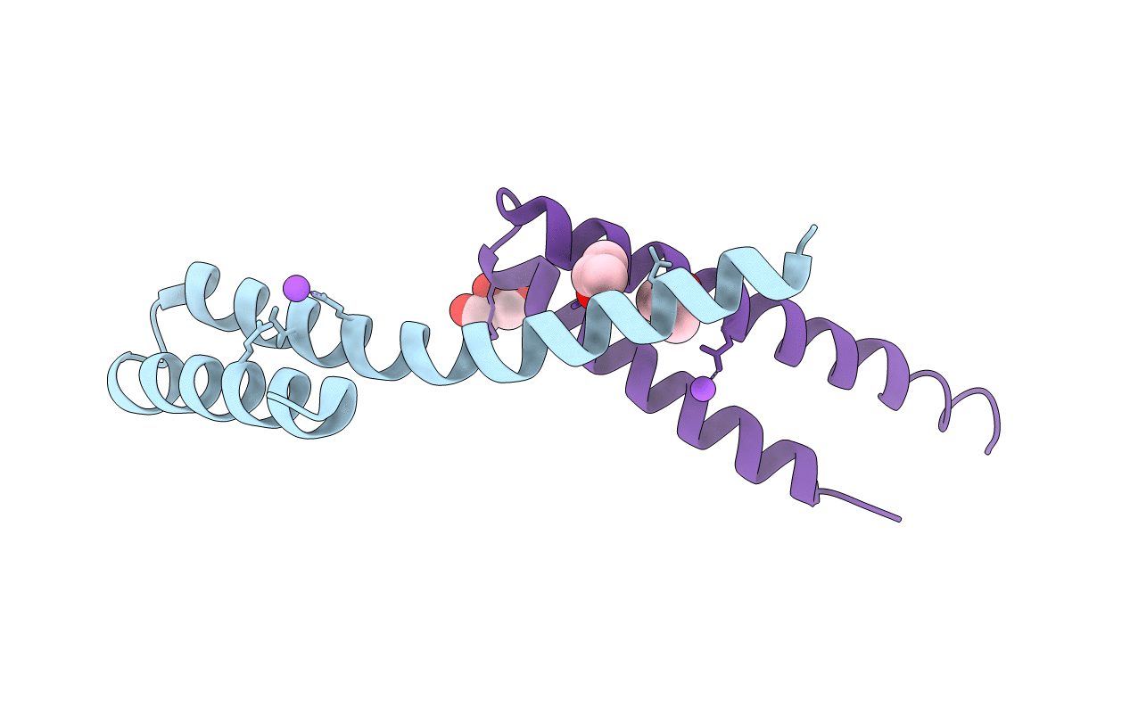
2CA5 image
Deposition Date 2005-12-16
Release Date 2006-08-07
Last Version Date 2024-05-08
Entry Detail
PDB ID:
2CA5
Title:
MxiH needle protein of Shigella Flexneri (monomeric form, residues 1- 78)
Biological Source:
Source Organism:
Host Organism:
Method Details:
Experimental Method:
Resolution:
2.10 Å
R-Value Work:
0.19
R-Value Observed:
0.19
Space Group:
C 1 2 1
Macromolecular Entities
Polymer Type:polypeptide(L)
Molecule:MXIH
Gene (Uniprot):sctF
Chain IDs:A, B
Chain Length:85
Number of Molecules:2
Biological Source:SHIGELLA FLEXNERI
Primary Citation
Molecular Model of a Type III Secretion System Needle: Implications for Host-Cell Sensing
Proc.Natl.Acad.Sci.USA 103 12529 ? (2006)
PMID: 16888041 DOI: 10.1073/PNAS.0602689103

Abstact

Type III secretion systems are essential virulence determinants for many Gram-negative bacterial pathogens. The type III secretion system consists of cytoplasmic, transmembrane, and extracellular domains. The extracellular domain is a hollow needle protruding above the bacterial surface and is held within a basal body that traverses both bacterial membranes. Effector proteins are translocated, via this external needle, directly into host cells, where they subvert normal cell functions to aid infection. Physical contact with host cells initiates secretion and leads to formation of a pore, thought to be contiguous with the needle channel, in the host-cell membrane. Here, we report the crystal structure of the Shigella flexneri needle subunit MxiH and a complete model for the needle assembly built into our three-dimensional EM reconstruction. The model, combined with mutagenesis data, reveals that signaling of host-cell contact is relayed through the needle via intersubunit contacts and suggests a mode of binding for a tip complex.

Legend

Protein

Chemical

Disease

Primary Citation of related structures