2C9I image
Deposition Date 2005-12-12
Release Date 2007-01-16
Last Version Date 2024-10-16
Entry Detail
PDB ID:
2C9I
Title:
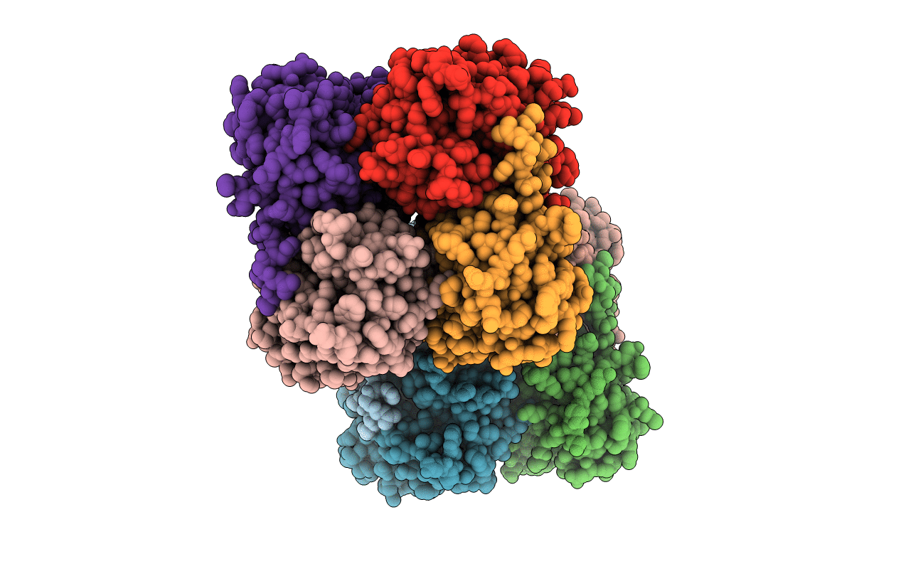
Structure of the fluorescent protein asFP499 from Anemonia sulcata
Biological Source:
Source Organism:
Host Organism:
Method Details:
Experimental Method:
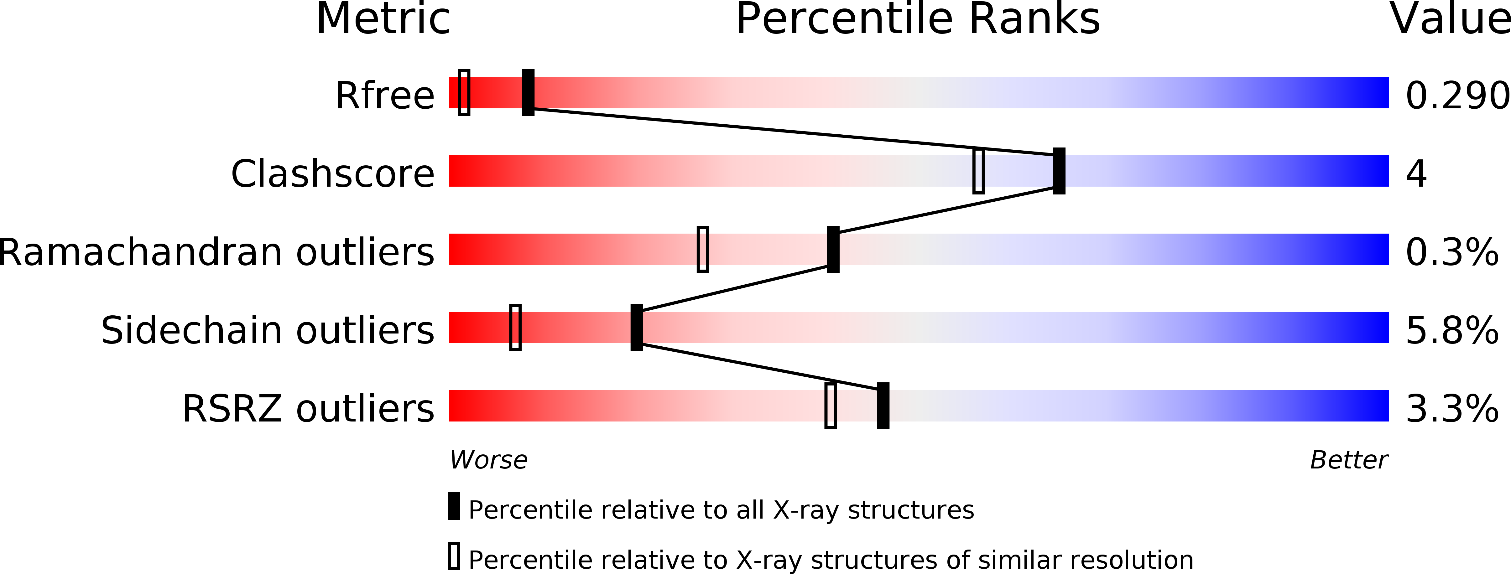
Resolution:
1.82 Å
R-Value Free:
0.29
R-Value Work:
0.24
R-Value Observed:
0.24
Space Group:
P 1 21 1
Macromolecular Entities
Structures with similar UniProt ID
Protein Blast
Polymer Type:polypeptide(L)
Molecule:GREEN FLUORESCENT PROTEIN ASFP499
Chain IDs:A, B, C, D, E, F, G, H
Chain Length:228
Number of Molecules:8
Biological Source:ANEMONIA SULCATA
Modified Residue
Compound ID Chain ID Parent Comp ID Details 2D Image
CRQ A GLN ?
Primary Citation
Chromophore-Protein Interactions in the Anthozoan Green Fluorescent Protein Asfp499
Biophys.J. 91 4210 ? (2006)
PMID: 16980366 DOI: 10.1529/BIOPHYSJ.106.087411

Abstact

Despite their similar fold topologies, anthozoan fluorescent proteins (FPs) can exhibit widely different optical properties, arising either from chemical modification of the chromophore itself or from specific interactions of the chromophore with the surrounding protein moiety. Here we present a structural and spectroscopic investigation of the green FP asFP499 from the sea anemone Anemonia sulcata var. rufescens to explore the effects of the protein environment on the chromophore. The optical absorption and fluorescence spectra reveal two discrete species populated in significant proportions over a wide pH range. Moreover, multiple protonation reactions are evident from the observed pH-dependent spectral changes. The x-ray structure of asFP499, determined by molecular replacement at a resolution of 1.85 A, shows the typical beta-barrel fold of the green FP from Aequorea victoria (avGFP). In its center, the chromophore, formed from the tripeptide Gln(63)-Tyr(64)-Gly(65), is tightly held by multiple hydrogen bonds in a polar cage that is structurally quite dissimilar to that of avGFP. The x-ray structure provides interesting clues as to how the spectroscopic properties are fine tuned by the chromophore environment.

Legend

Protein

Chemical

Disease