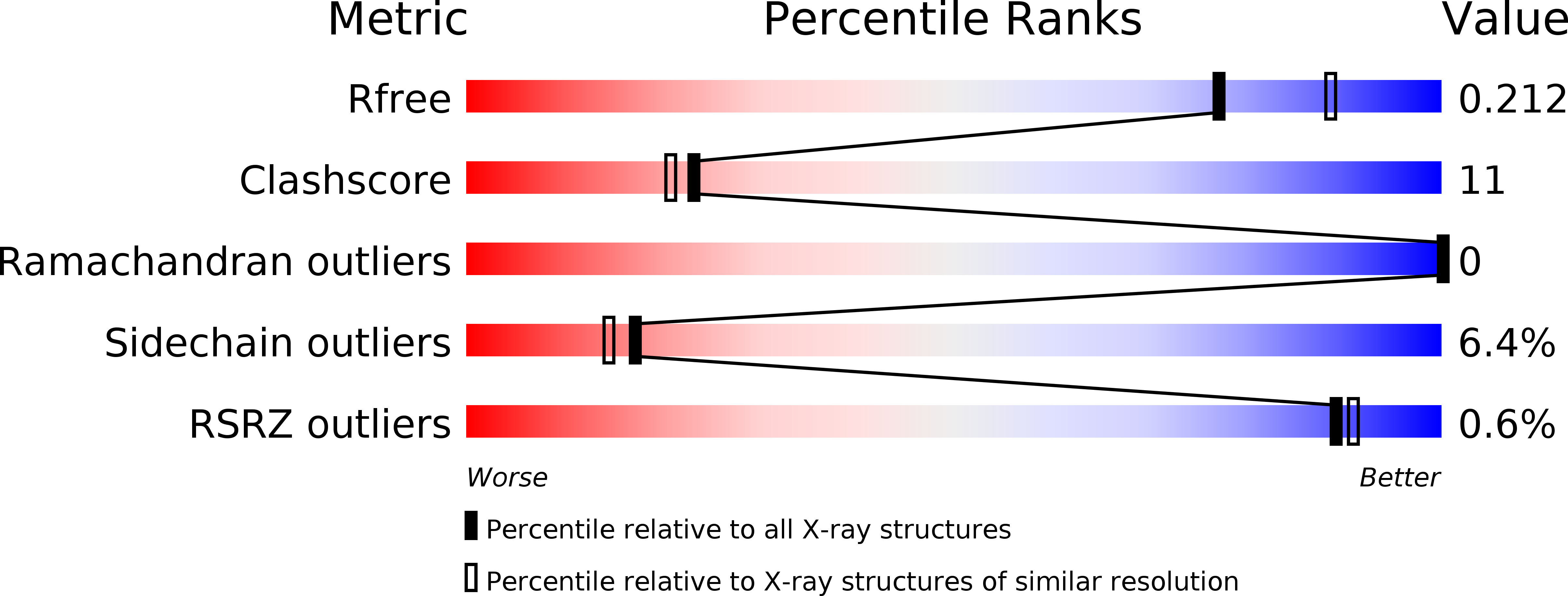
2C9E image
Entry Detail
PDB ID:
2C9E
Keywords:
Title:
Peridinin-chlorophyll a protein, high-salt form
Biological Source:
Source Organism:
PDB Version:
Deposition Date:
2005-12-09
Release Date:
2007-02-20
Method Details:
Experimental Method:
Resolution:
2.10 Å
R-Value Free:
0.21
R-Value Work:
0.17
R-Value Observed:
0.17
Space Group:
P 1 21 1
Macromolecular Entities
Polymer Type:polypeptide(L)
Description:PERIDININ-CHLOROPHYLL A PROTEIN
Chain IDs:A
Chain Length:327
Number of Molecules:1
Biological Source:AMPHIDINIUM CARTERAE
Primary Citation
X-Ray Structure of the High-Salt Form of the Peridinin-Chlorophyll A-Protein from the Dinoflagellate Amphidinium Carterae: Modulation of the Spectral Properties of Pigments by the Protein Environment.
Biochemistry 48 4466 ? (2009)
PMID: 19371099 DOI: 10.1021/BI802320Q

Abstact

Light-harvesting complexes have evolved into very different structures but fulfill the same function, efficient harvesting of solar energy. In these complexes, pigments are fine-tuned and properly arranged to gather incoming photons. In the photosynthetic dinoflagellate Amphidinium carterae, two variants of the soluble light-harvesting complex PCP have been found [main form PCP (MFPCP) and high-salt PCP (HSPCP)], which show small variations in their pigment arrangement and tuning mechanisms. This feature makes them ideal models for studying pigment-protein interactions. Here we present the X-ray structure of the monomeric HSPCP determined at 2.1 A resolution and compare it to the structure of trimeric MFPCP. Despite the high degree of structural similarity (rmsd C(alpha)-C(alpha) of 1.89 A), the sequence variations lead to a changed overall pigment composition which includes the loss of two carotenoid molecules and a dramatic rearrangement of the chlorophyll phytol chains and of internal lipid molecules. On the basis of a detailed structural comparison, we favor a macrocycle geometry distortion of the chlorophylls rather than an electrostatic effect to explain energetic splitting of the chlorophyll a Q(y) bands [Ilagan, R. P. (2006) Biochemistry 45, 14052-14063]. Our analysis supports their assignment of peridinin 611* as the single blue-shifted peridinin in HSPCP but also highlights another electrostatic feature due to glutamate 202 which could add to the observed binding site asymmetry of the 611*/621* peridinin pair.

Legend

Protein

Chemical

Disease

Primary Citation of related structures