2C8I image
Deposition Date 2005-12-05
Release Date 2006-01-17
Last Version Date 2024-05-08
Entry Detail
PDB ID:
2C8I
Keywords:
Title:
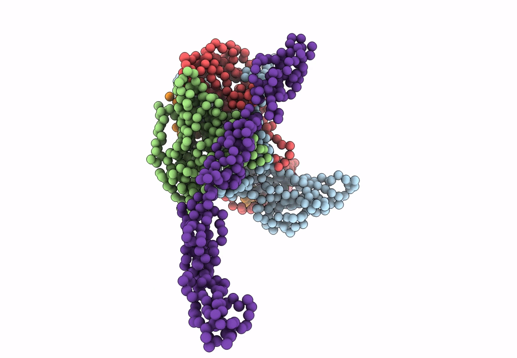
Complex Of Echovirus Type 12 With Domains 1, 2, 3 and 4 Of Its Receptor Decay Accelerating Factor (Cd55) By Cryo Electron Microscopy At 16 A
Biological Source:
Source Organism:
HOMO SAPIENS (Taxon ID: 9606)
HUMAN ECHOVIRUS 11 (Taxon ID: 12078)
Host Organism:
Method Details:
Experimental Method:
Resolution:
14.00 Å
Aggregation State:
PARTICLE
Reconstruction Method:
SINGLE PARTICLE
Macromolecular Entities
Polymer Type:polypeptide(L)
Molecule:ECHOVIRUS 11 COAT PROTEIN VP1
Chain IDs:A
Chain Length:289
Number of Molecules:1
Biological Source:HUMAN ECHOVIRUS 11
Polymer Type:polypeptide(L)
Molecule:ECHOVIRUS 11 COAT PROTEIN VP2
Chain IDs:B
Chain Length:252
Number of Molecules:1
Biological Source:HUMAN ECHOVIRUS 11
Polymer Type:polypeptide(L)
Molecule:ECHOVIRUS 11 COAT PROTEIN VP3
Chain IDs:C
Chain Length:238
Number of Molecules:1
Biological Source:HUMAN ECHOVIRUS 11
Polymer Type:polypeptide(L)
Molecule:ECHOVIRUS 11 COAT PROTEIN VP4
Chain IDs:D
Chain Length:60
Number of Molecules:1
Biological Source:HUMAN ECHOVIRUS 11
Polymer Type:polypeptide(L)
Molecule:COMPLEMENT DECAY-ACCELERATING FACTOR
Gene (Uniprot):CD55
Chain IDs:E
Chain Length:316
Number of Molecules:1
Biological Source:HOMO SAPIENS
Ligand Molecules
Primary Citation
Structural and Functional Insights Into the Interaction of Echoviruses and Decay-Accelerating Factor.
J.Biol.Chem. 281 5169 ? (2006)
PMID: 16272562 DOI: 10.1074/JBC.M510362200

Abstact

Many enteroviruses bind to the complement control protein decay-accelerating factor (DAF) to facilitate cell entry. We present here a structure for echovirus (EV) type 12 bound to DAF using cryo-negative stain transmission electron microscopy and three-dimensional image reconstruction to 16-A resolution, which we interpreted using the atomic structures of EV11 and DAF. DAF binds to a hypervariable region of the capsid close to the 2-fold symmetry axes in an interaction that involves mostly the short consensus repeat 3 domain of DAF and the capsid protein VP2. A bulge in the density for the short consensus repeat 3 domain suggests that a loop at residues 174-180 rearranges to prevent steric collision between closely packed molecules at the 2-fold symmetry axes. Detailed analysis of receptor interactions between a variety of echoviruses and DAF using surface plasmon resonance and comparison of this structure (and our previous work; Bhella, D., Goodfellow, I. G., Roversi, P., Pettigrew, D., Chaudhry, Y., Evans, D. J., and Lea, S. M. (2004) J. Biol. Chem. 279, 8325-8332) with reconstructions published for EV7 bound to DAF support major differences in receptor recognition among these viruses. However, comparison of the electron density for the two virus.receptor complexes (rather than comparisons of the pseudo-atomic models derived from fitting the coordinates into these densities) suggests that the dramatic differences in interaction affinities/specificities may arise from relatively subtle structural differences rather than from large-scale repositioning of the receptor with respect to the virus surface.

Legend

Protein

Chemical

Disease

Primary Citation of related structures