2C7Z image
Entry Detail
PDB ID:
2C7Z
Keywords:
Title:
Plant enzyme crystal form II
Biological Source:
Source Organism:
Host Organism:
PDB Version:
Deposition Date:
2005-11-30
Release Date:
2006-05-17
Method Details:
Experimental Method:
Resolution:
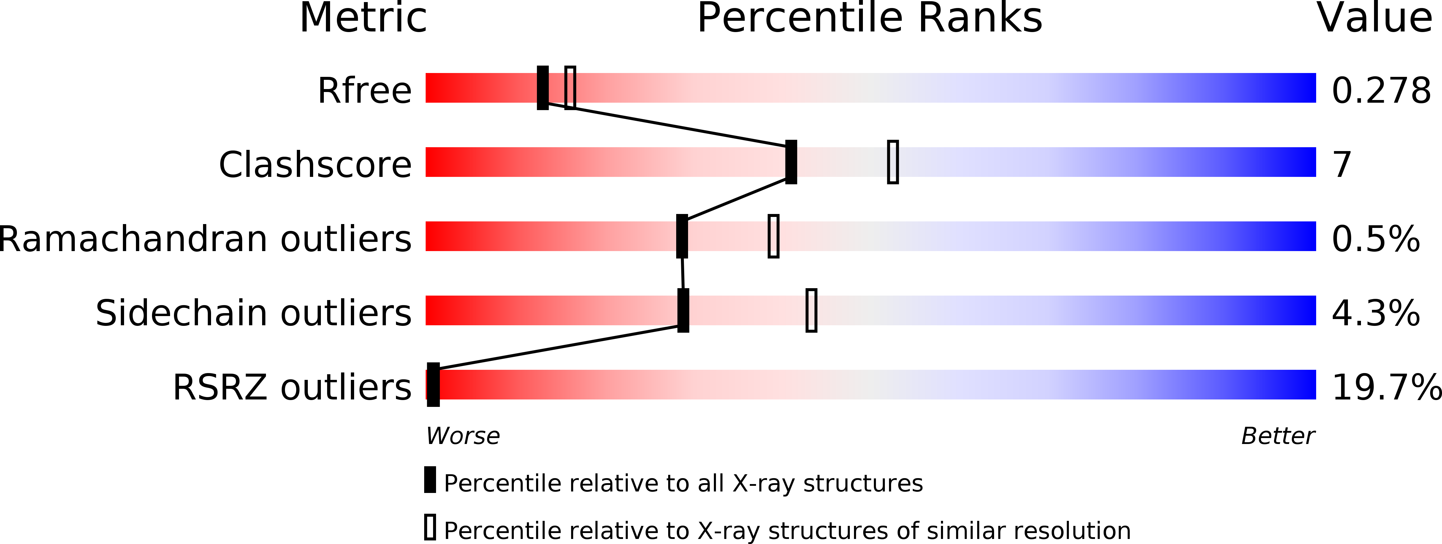
2.37 Å
R-Value Free:
0.27
R-Value Work:
0.19
R-Value Observed:
0.20
Space Group:
P 21 21 2
Macromolecular Entities
Polymer Type:polypeptide(L)
Description:3-KETOACYL-COA THIOLASE 2
Chain IDs:A
Chain Length:404
Number of Molecules:1
Biological Source:ARABIDOPSIS THALIANA
Primary Citation
The Crystal Structure of a Plant 3-Ketoacyl-Coa Thiolase Reveals the Potential for Redox Control of Peroxisomal Fatty Acid Beta-Oxidation.
J.Mol.Biol. 359 347 ? (2006)
PMID: 16630629 DOI: 10.1016/J.JMB.2006.03.032

Abstact

Crystal structures of peroxisomal Arabidopsis thaliana 3-ketoacyl-CoA thiolase (AtKAT), an enzyme of fatty acid beta-oxidation, are reported. The subunit, a typical thiolase, is a combination of two similar alpha/beta domains capped with a loop domain. The comparison of AtKAT with the Saccharomyces cerevisiae homologue (ScKAT) structure reveals a different placement of subunits within the functional dimers and that a polypeptide segment forming an extended loop around the open catalytic pocket of ScKAT converts to alpha-helix in AtKAT, and occludes the active site. A disulfide is formed between Cys192, on this helix, and Cys138, a catalytic residue. Access to Cys138 is determined by the structure of this polypeptide segment. AtKAT represents an oxidized, previously unknown inactive form, whilst ScKAT is the reduced and active enzyme. A high level of sequence conservation is observed, including Cys192, in eukaryotic peroxisomal, but not mitochondrial or prokaryotic KAT sequences, for this labile loop/helix segment. This indicates that KAT activity in peroxisomes is influenced by a disulfide/dithiol change linking fatty acid beta-oxidation with redox regulation.

Legend

Protein

Chemical

Disease

Primary Citation of related structures