2C74 image
Deposition Date 2005-11-17
Release Date 2005-12-02
Last Version Date 2024-11-20
Entry Detail
PDB ID:
2C74
Title:
14-3-3 Protein Eta (Human) Complexed to Peptide
Biological Source:
Source Organism:
Method Details:
Experimental Method:
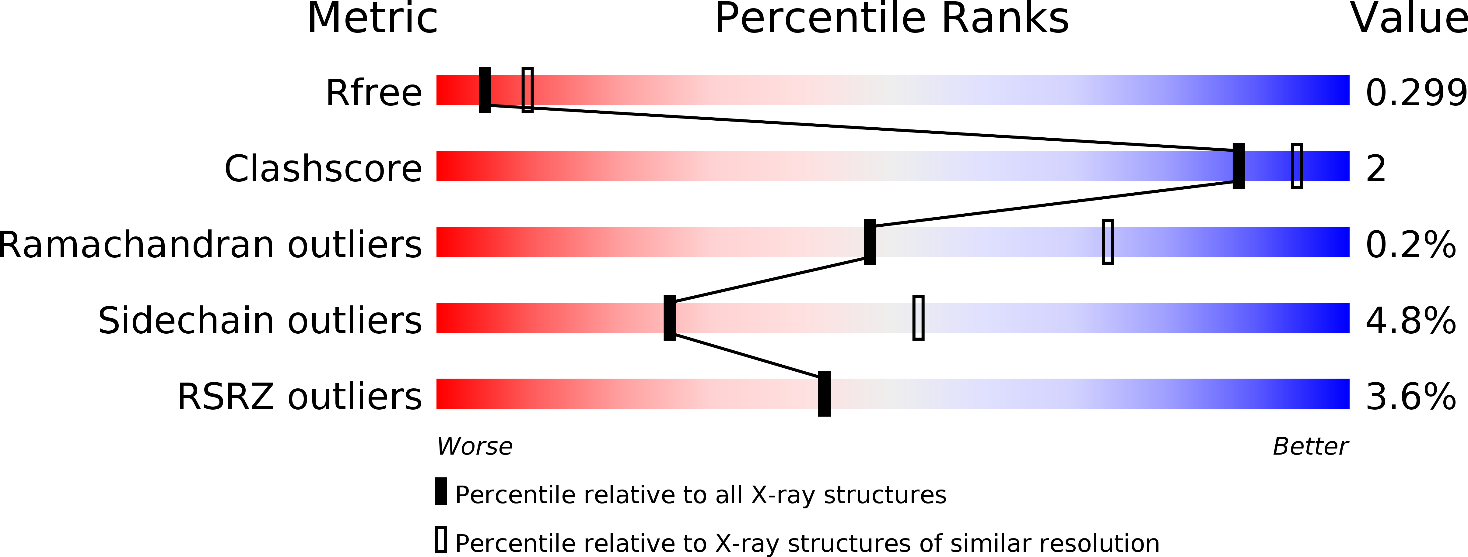
Resolution:
2.70 Å
R-Value Free:
0.29
R-Value Work:
0.22
R-Value Observed:
0.22
Space Group:
P 21 21 21
Macromolecular Entities
Structures with similar UniProt ID
Protein Blast
Polymer Type:polypeptide(L)
Molecule:14-3-3 PROTEIN ETA
Gene (Uniprot):YWHAH
Chain IDs:A, B
Chain Length:247
Number of Molecules:2
Biological Source:HOMO SAPIENS
Polymer Type:polypeptide(L)
Molecule:CONSENSUS PEPTIDE MODE 1 FOR 14-3-3 PROTEINS
Chain IDs:C (auth: P), D (auth: Q)
Chain Length:7
Number of Molecules:2
Biological Source:synthetic construct
Modified Residue
Compound ID Chain ID Parent Comp ID Details 2D Image
SEP C SER PHOSPHOSERINE
Ligand Molecules
Primary Citation
Structural basis for protein-protein interactions in the 14-3-3 protein family.
Proc. Natl. Acad. Sci. U.S.A. 103 17237 17242 (2006)
PMID: 17085597 DOI: 10.1073/pnas.0605779103

Abstact

The seven members of the human 14-3-3 protein family regulate a diverse range of cell signaling pathways by formation of protein-protein complexes with signaling proteins that contain phosphorylated Ser/Thr residues within specific sequence motifs. Previously, crystal structures of three 14-3-3 isoforms (zeta, sigma, and tau) have been reported, with structural data for two isoforms deposited in the Protein Data Bank (zeta and sigma). In this study, we provide structural detail for five 14-3-3 isoforms bound to ligands, providing structural coverage for all isoforms of a human protein family. A comparative structural analysis of the seven 14-3-3 proteins revealed specificity determinants for binding of phosphopeptides in a specific orientation, target domain interaction surfaces and flexible adaptation of 14-3-3 proteins through domain movements. Specifically, the structures of the beta isoform in its apo and peptide bound forms showed that its binding site can exhibit structural flexibility to facilitate binding of its protein and peptide partners. In addition, the complex of 14-3-3 beta with the exoenzyme S peptide displayed a secondary structural element in the 14-3-3 peptide binding groove. These results show that the 14-3-3 proteins are adaptable structures in which internal flexibility is likely to facilitate recognition and binding of their interaction partners.

Legend

Protein

Chemical

Disease

Primary Citation of related structures