
Deposition Date
2005-11-02
Release Date
2005-12-07
Last Version Date
2024-11-13
Entry Detail
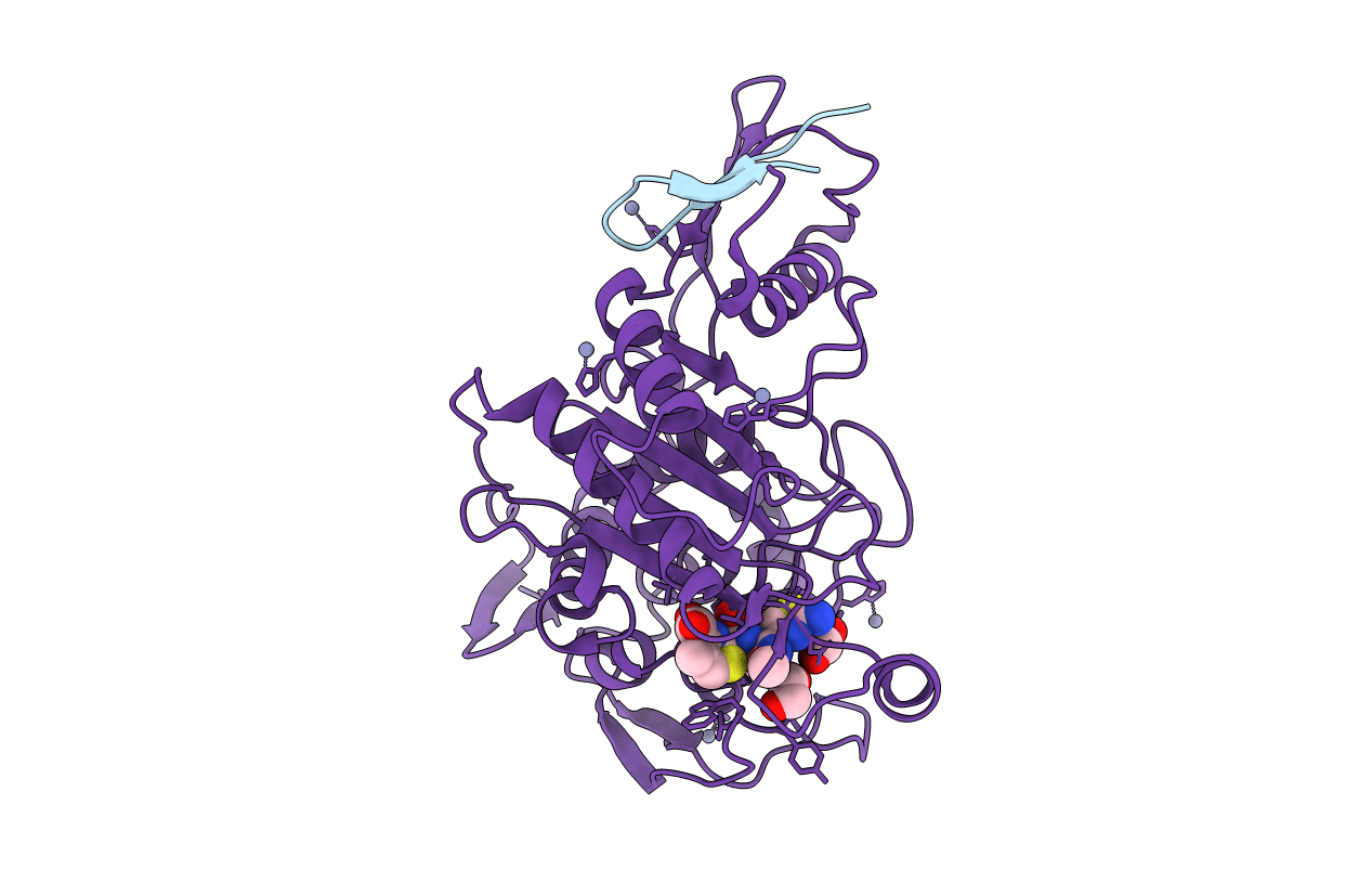
PDB ID:
2C5W
Keywords:
Title:
PENICILLIN-BINDING PROTEIN 1A (PBP-1A) ACYL-ENZYME COMPLEX (CEFOTAXIME) FROM STREPTOCOCCUS PNEUMONIAE
Biological Source:
Source Organism(s):
STREPTOCOCCUS PNEUMONIAE (Taxon ID: 171101)
Expression System(s):
Method Details:
Experimental Method:
Resolution:
2.55 Å
R-Value Free:
0.25
R-Value Work:
0.23
R-Value Observed:
0.23
Space Group:
C 2 2 21
