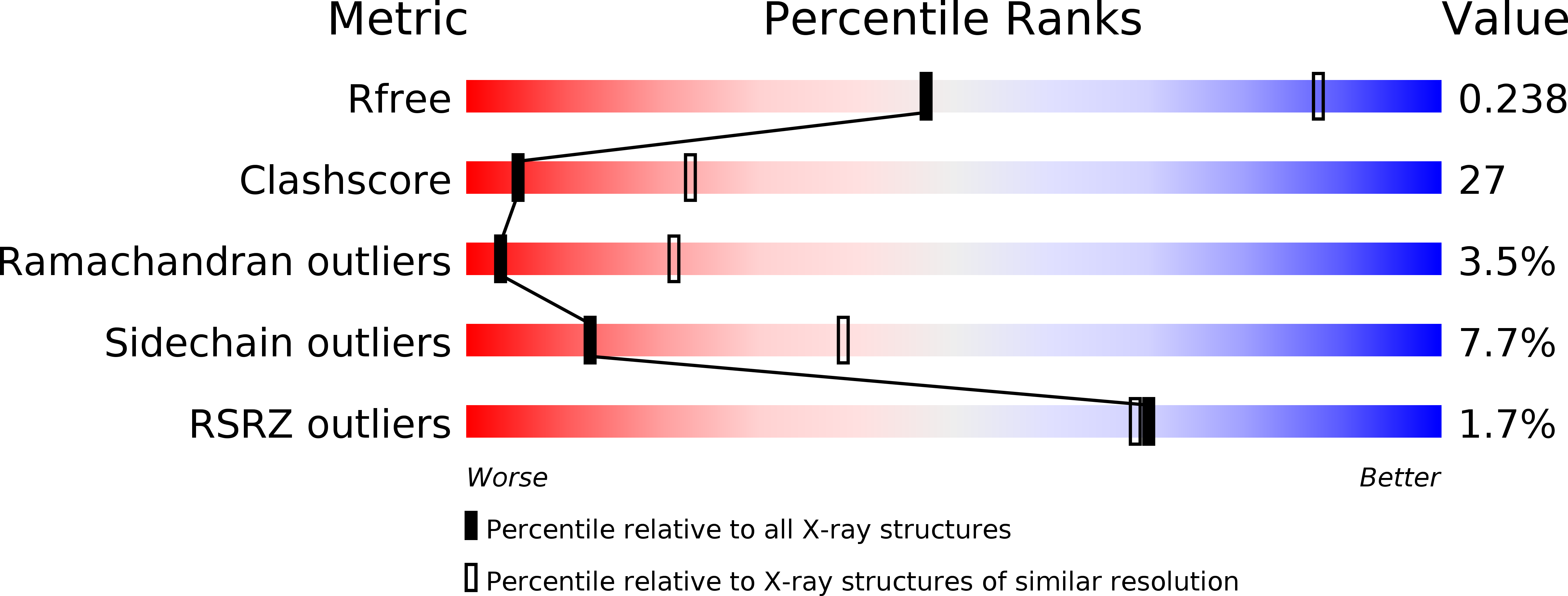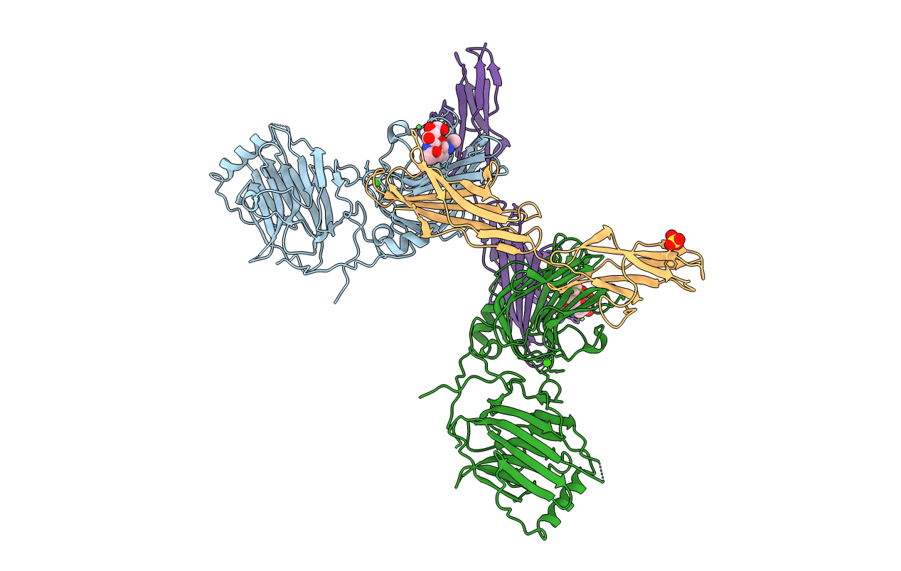
2C5D image
Deposition Date 2005-10-26
Release Date 2005-12-19
Last Version Date 2024-11-20
Entry Detail
PDB ID:
2C5D
Title:
Structure of a minimal Gas6-Axl complex
Biological Source:
Source Organism:
HOMO SAPIENS (Taxon ID: 9606)
Host Organism:
Method Details:
Experimental Method:
Resolution:
3.30 Å
R-Value Free:
0.26
R-Value Work:
0.24
R-Value Observed:
0.24
Space Group:
P 61
Macromolecular Entities
Polymer Type:polypeptide(L)
Molecule:GROWTH-ARREST-SPECIFIC PROTEIN 6 PRECURSOR
Gene (Uniprot):GAS6
Chain IDs:A, B
Chain Length:422
Number of Molecules:2
Biological Source:HOMO SAPIENS
Polymer Type:polypeptide(L)
Molecule:TYROSINE-PROTEIN KINASE RECEPTOR UFO
Gene (Uniprot):AXL
Chain IDs:C, D
Chain Length:195
Number of Molecules:2
Biological Source:HOMO SAPIENS
Modified Residue
Compound ID Chain ID Parent Comp ID Details 2D Image
ASN A ASN GLYCOSYLATION SITE
Primary Citation

Abstact

Receptor tyrosine kinases of the Axl family are activated by the vitamin K-dependent protein Gas6. Axl signalling plays important roles in cancer, spermatogenesis, immunity, and platelet function. The crystal structure at 3.3 A resolution of a minimal human Gas6/Axl complex reveals an assembly of 2:2 stoichiometry, in which the two immunoglobulin-like domains of the Axl ectodomain are crosslinked by the first laminin G-like domain of Gas6, with no direct Axl/Axl or Gas6/Gas6 contacts. There are two distinct Gas6/Axl contacts of very different size, both featuring interactions between edge beta-strands. Structure-based mutagenesis, protein binding assays and receptor activation experiments demonstrate that both the major and minor Gas6 binding sites are required for productive transmembrane signalling. Gas6-mediated Axl dimerisation is likely to occur in two steps, with a high-affinity 1:1 Gas6/Axl complex forming first. Only the minor Gas6 binding site is highly conserved in the other Axl family receptors, Sky/Tyro3 and Mer. Specificity at the major contact is suggested to result from the segregation of charged and apolar residues to opposite faces of the newly formed beta-sheet.

Legend

Protein

Chemical

Disease

Primary Citation of related structures